Figures & data
Figure 1 Schematic of the anatomical and non-anatomical causes of OSA.
Notes: Some degree of anatomical vulnerability is present in OSA. However, the extent of impairment varies widely between patients. The non-anatomical contributors, which are present in approximately 70% of OSA patients, play a key role in mediating the absence or presence of OSA. In the schematic, the gray tracings indicate the desired response, whereas the black tracings represent impairment in the non-anatomical trait. Refer to the text for further detail. Reprinted from Chest, Carberry JC, Amatoury J, Eckert DJ, Personalized management approach for OSA, Epub 2017 June 16, Copyright (2017), with permission from Elsevier.Citation36
Abbreviations: EEG, electroencephalography; EMG, genioglossus electromyography; MTA, 100 ms moving time average of the rectified raw EMG signal; OSA, obstructive sleep apnea.
Abbreviations: EEG, electroencephalography; EMG, genioglossus electromyography; MTA, 100 ms moving time average of the rectified raw EMG signal; OSA, obstructive sleep apnea.
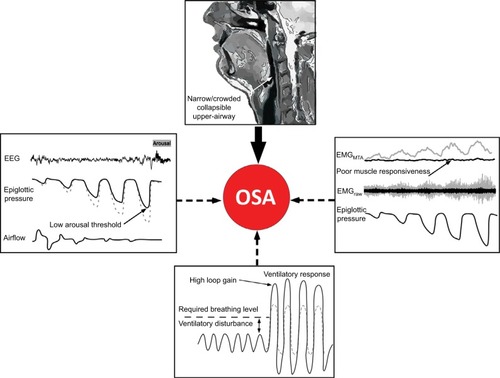
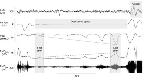
Figure 2 Example of minimal genioglossus muscle responsiveness.
Notes: In this example of a naturally occurring apnea, despite clear phasic activation of the genioglossus muscle (as shown in the raw and MTA genioglossus EMG channels), there is minimal activation of genioglossus during the respiratory event. This is despite substantial increasing negative epiglottic pressure (Pepi) swings from the first to last effort (last effort nadir epiglottic pressure = arousal threshold). It is only when cortical arousal occurs (as shown in the EEG channel) that major genioglossus activation occurs (signal clipped in this example) and airflow is restored.
Abbreviations: EEG, electroencephalography; EMG, genioglossus electromyography; MTA, 100 ms moving time average of the rectified raw EMG signal.
Abbreviations: EEG, electroencephalography; EMG, genioglossus electromyography; MTA, 100 ms moving time average of the rectified raw EMG signal.
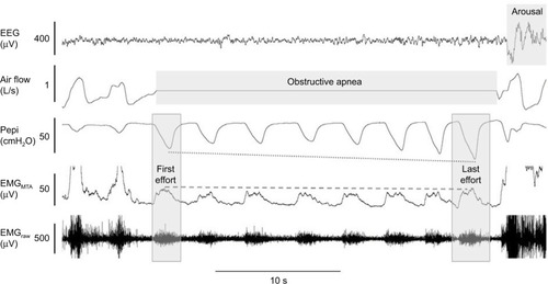
Figure 3 Example of robust genioglossus muscle responsiveness and restoration of airflow without cortical arousal.
Notes: In contrast to the example in , in this example of a naturally occurring hypopnea, there is robust activation of the genioglossus muscle (as shown in the raw and MTA genioglossus EMG channels), to increasing negative epiglottic pressure (Pepi) swings from the first to last effort which ultimately results in recovery of airflow without cortical arousal.
Abbreviations: EEG, electroencephalogram; EMG, genioglossus electromyography; MTA, 100 ms moving time average of the rectified raw EMG signal.
Abbreviations: EEG, electroencephalogram; EMG, genioglossus electromyography; MTA, 100 ms moving time average of the rectified raw EMG signal.
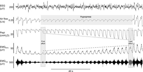
Figure 4 Example of robust genioglossus muscle responsiveness without restoration of airflow.
Notes: In contrast to the example in , in this example of a naturally occurring apnea, there is robust activation of the genioglossus muscle (as shown in the raw and MTA genioglossus EMG channels) to increasing negative epiglottic pressure (Pepi) swings from the first to last effort (last effort nadir epiglottic pressure = arousal threshold). However, despite substantial genioglossus muscle activation during the apnea, it is insufficient to restore airflow, which only occurs with cortical arousal (as shown in the EEG channel). The genioglossus signal is clipped in this example when airflow is restored with arousal.
Abbreviations: EEG, electroencephalogram; EMG, genioglossus electromyography; MTA, 100 ms moving time average of the rectified raw EMG signal.
Abbreviations: EEG, electroencephalogram; EMG, genioglossus electromyography; MTA, 100 ms moving time average of the rectified raw EMG signal.