
Figures & data
Table 1 Baseline Characteristics of the Study Population
Table 2 Imaging Parameters of the Study Population
Table 3 Clinical Variables Associated with LGE% from Univariate and Multivariate Analysis
Figure 1 Representative LGE and non-sustained ventricular tachycardia image from patients. (A, B) The 4-chamber long-axis and left ventricular short-axis image from a patient who have severe OSA with a value of 13.99%LGE (arrow); (C) the representative non-sustained ventricular tachycardia image from the same patient; (D–E) the 4-chamber long-axis and left ventricular short-axis image from a patient who have severe OSA with a value of 21.88%LGE (arrow).
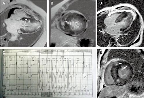
Table 4 Relationship Between Arrhythmias and OSA
Figure 2 The value of late gadolinium enhancement according to the presence of various arrhythmias between patients with and without arrhythmias. (A) Premature ventricular contractions; (B) paired premature ventricular contractions; (C) ventricular bigeminy; (D) ventricular trigeminy; (E) premature atrial contractions; (F) atrial bigeminy; (G) non-sustained ventricular tachycardia; (H) supraventricular tachycardia.
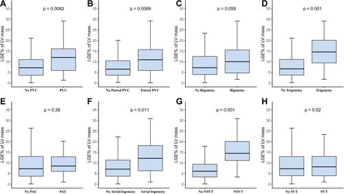
Table 5 Adjusted Logistic Regression Among Arrhythmias and Significant Variables from Univariate Analysis