
Figures & data
Figure 1 Flow chart of the study. From January 2018 to December 2019, 200 physical examinees (including 108 men and 92 women) aged 18–60 years admitted for MRI and overnight polysomnography (PSG) at the sleep unit of the Second Affiliated Hospital of Zhengzhou University were enrolled for the present analysis. During the night of PS-G, participants were under supervision of a qualified technician and underwent PSG monitoring for at least 7 h. Among the 200 subjects enrolled into the present work, two were excluded because of the low MRI scan quality, whereas 61 were ruled out from this work due to diabetes, hypertension, cardiovascular disease or neurological disorder. In the diagnosis process of PSG, six subjects with the overall sleep time <4 h were eliminated from this work. In addition, 23 smokers or drinkers and another one meeting the criteria of current major depression were also eliminated. Therefore, finally 107 subjects (including 44 (41.12%) females) were enrolled, with the age of (31.72±11.86) years. 44 (41.12%) subjects showing the AHI<5/h were enrolled as the control group; 25 (23.36%) had AHI of 5–15/h (mild); 20 (18.69%) had AHI of 15–30/h (moderate); and 18 (16.82%) had AHI >30/h (severe).
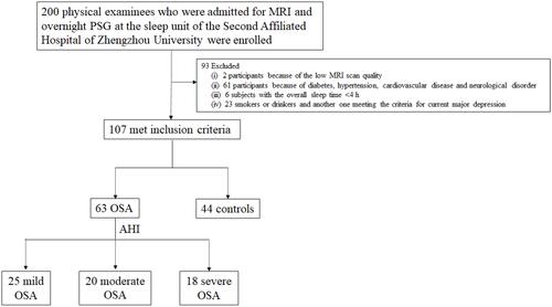
Figure 2 Selection of PVS observation layer shown in the sagittal view. (i) frontoparietal subcortical white matter layer; (ii) centrum semiovale layer; (iii) basal ganglia layer.
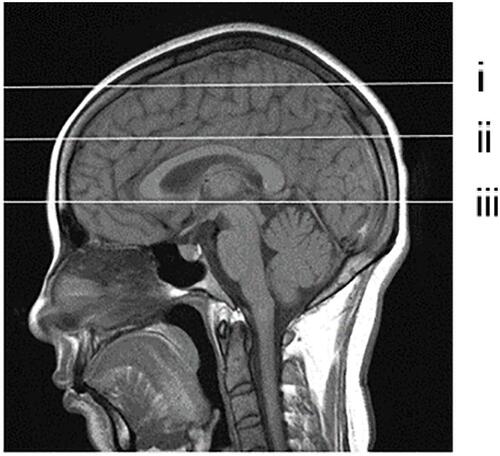
Table 1 Demographic Features and EPVS in Control and OSA Groups
Table 2 Analysis of PSG Results Among All Groups
Figure 3 EPVS in OSA group observed at different layers. (A–C) display the frontoparietal subcortical white matter layer, centrum semiovale layer and basal ganglia layer; EPVS (white arrows).
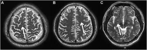
Table 3 Partial Spearman’s Rank Order Correlation Results Unadjusted for Covariates
Table 4 Partial Spearman’s Rank Order Correlation Results Controlling for Age, BMI, ESS