Figures & data
Figure 1 Schematic diagram of OSA patient collection in this study.
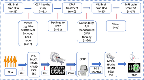
Table 1 Demographic and Clinical Data of Pre-CPAP and Post-CPAP
Table 2 Distribution of MD Differences in OSA Patients After 12 Months of CPAP Treatment
Table 3 Distribution of RD Differences in OSA Patients After 12 Months of CPAP Treatment
Figure 2 TBSS analysis of white matter fibres in OSA patients before and after CPAP treatment, green: mean fractional anisotropy (FA) skeleton; blue and red: area of change of DTI values in the OSA group after CPAP treatment (MD and RD), p<0.05.
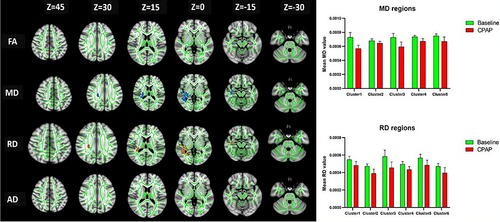
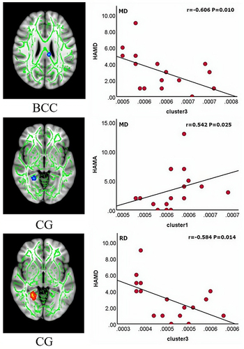
Figure 3 Correlation of MD and RD values with clinical assessment scales in OSA patients after treatment.
Abbreviations: BCC, body of corpus callosum; CG, Cingulum.