Figures & data
Figure 1 ROIs used in quantitative analysis of DNs in coronal sections of the rat brain.
Notes: (A) Schematic of a coronal section through the brain at AP -2 mm relative to bregma (adapted from Ref Citation57). □ indicate ROIs (600 µm ×400 µm) used for cell counts in the Co, SON, and in the Pin. (B) SON (×100 magnification), illustrating generalized hyperbasophilia and the absence of obvious compacted DN. (C) Photomicrograph of cortical neurons in a pin-prick positive control at ×100 magnification. DNs (basophilic with a compacted triangular shape) are visible in the tissue adjacent to the pin lesion. Tissues stained with toluidine blue. Scale bars, 100 µm.
Abbreviations: ROI, region of interest; DN, dark neuron; Co, cortex; SON, supraoptic nucleus; Pin, cortex pin-prick perilesion.
Abbreviations: ROI, region of interest; DN, dark neuron; Co, cortex; SON, supraoptic nucleus; Pin, cortex pin-prick perilesion.
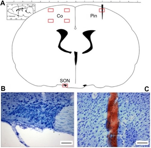
Figure 2 Artifactual DN formation in the cortex following immersion fixation.
Notes: (A) Photomicrograph (×100) of a representative perfusion-fixed cortical region of interest (HCC group) demonstrating the absence of DN. (B) Photomicrograph (×100) of a representative immersion-fixed cortical region of interest (IMM group), showing numerous DNs (⇨heads) scattered at random among normal neurons. Stained with toluidine blue. Scale bars, 50 µm.
Abbreviations: DN, dark neuron; HCC, perfusion-fixed home cage control; IMM, immersion-fixed positive control.
Abbreviations: DN, dark neuron; HCC, perfusion-fixed home cage control; IMM, immersion-fixed positive control.
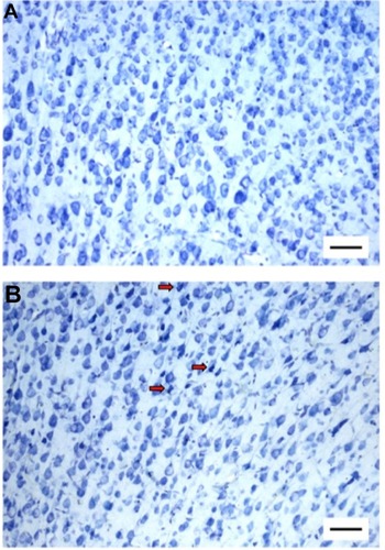
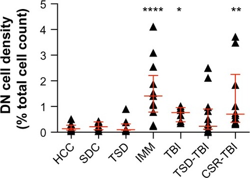
Figure 3 DN counts in rat sensorimotor cortex.
Notes: DN density is expressed as % of total cell count in the combined cortex regions of interest. Symbols indicate individual animals, error bars (Image) denote median ± interquartile range, and asterisks indicate a statistically significant difference vs HCC (Kruskal–Wallis test followed by Dunn’s multiple comparisons test: *P<0.05, **P<0.01, and ****P<0.0001).
Abbreviations: DN, dark neuron; HCC, perfusion-fixed home cage control; SDC, perfusion-fixed stimulus control; TSD, total sleep deprivation; IMM, immersion-fixed positive control; TBI, traumatic brain injury; TSD-TBI, TSD followed by TBI; CSR-TBI, chronic sleep restriction followed by TBI.
Abbreviations: DN, dark neuron; HCC, perfusion-fixed home cage control; SDC, perfusion-fixed stimulus control; TSD, total sleep deprivation; IMM, immersion-fixed positive control; TBI, traumatic brain injury; TSD-TBI, TSD followed by TBI; CSR-TBI, chronic sleep restriction followed by TBI.