
Figures & data
Figure 1 Schematic of the implant and delivery system.
Notes: The implant pictured has single-hole loop attachment points (A). The deployed implant is depicted in the tongue base (B) and soft palate (C), with the implant tool in situ for visualization of the delivery method).
Abbreviation: PLG, polylactide-co-glycolide.
Abbreviation: PLG, polylactide-co-glycolide.
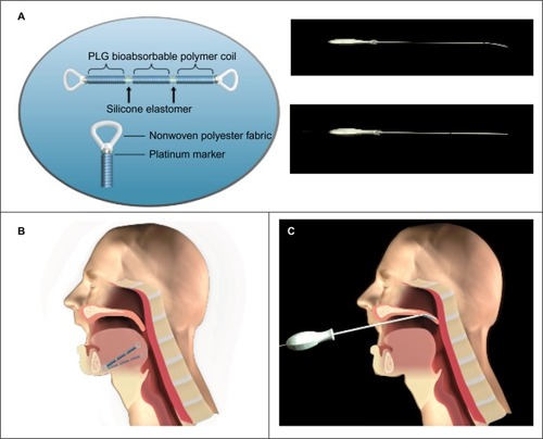
Figure 2 FEA model of the palate and tongue (A) and single-tongue implant (B).
Abbreviation: FEA, finite element analysis.
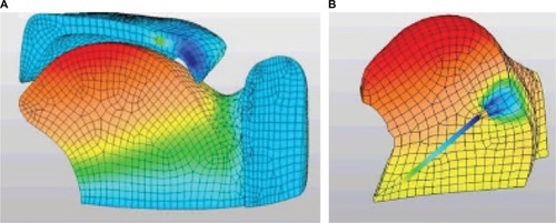
Figure 3 685207-F1 pre- and postfatigue testing static conditioning test
Note: The dynamic modulus of the implant was not affected by 5,260,000 stretch cycles; values before (red) and after (blue) stretching were virtually identical.
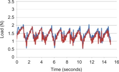
Figure 4 Exemplar histological samples: inflammatory response at the attachment points and middle of the linear component of the implants of sheep at 30 days and 60 days.
Notes: (A) Fibrous tissue fills the center hole of this device (center of image), bridging between muscle at the top and bottom of image. The two circular voids represent each side of the attachment point loop at 30 days. (B) This micrograph shows the typical 30-day reaction with the fast-eroding PLG; note that a residual PLG layer of ~0.5 mm surrounds the device (void in center of image). There is mild accompanying foreign body-type inflammation directed at the PLG material, with a further encircling thin band of fibrous connective tissue. (C) There is mature fibrous tissue spanning the attachment opening and minimal inflammation at 60 days postimplantation. (D) There is a very thin circular layer of bland fibrous tissue encapsulating the contractile element at 60 days. No residual PLG is present and inflammation is insignificant.
Abbreviation: PLG, polylactide-co-glycolide.
Abbreviation: PLG, polylactide-co-glycolide.
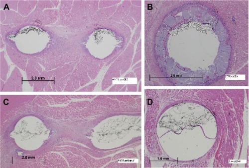
Table 1 Single-hole silicone loop and paddle-shaped implant ends resulted in the best fibrous tissue attachment in lumbar muscle tissue in the ovine model
Table 2 For both tongue and soft palate implants in the canine model, there was little inflammatory response for either silicone durometer tested