Figures & data
Figure 1 CT venogram axial view shows compression of the left iliac vein (blue arrow) by the right iliac artery (red arrow) at the level of the 4th lumbar vertebra (L4).
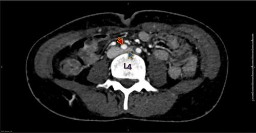
Figure 2 CT venogram coronal view shows compression of the left iliac vein (blue arrow) by the right iliac artery (red arrow).
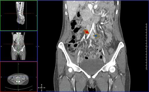
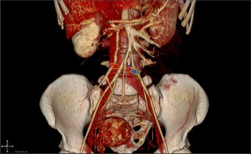
Figure 3 3D reconstruction of the CT venogram shows compression of the left iliac vein (blue arrow) by the right iliac artery (red arrow) at the level of the 4th lumbar vertebra (L4).