
Figures & data
Figure 1 CT scan of the neck shows multiple peripheral contrast-enhancing abscesses in both sides of the neck (bilateral neck abscess).
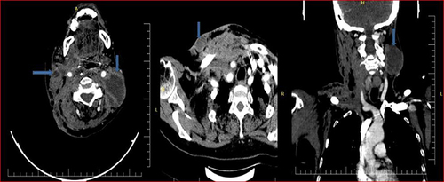
Figure 2 Coronal and axial neck CT scan reveals inflamed soft tissue with loculated gas in the right side of the neck and supraclavicular region (necrotising fasciitis of the neck).
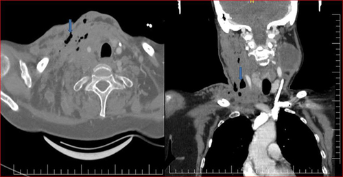
Figure 3 Axial chest CT shows several peripheral predominant nodules and some cavitation in both lungs (septic pulmonary embolism).
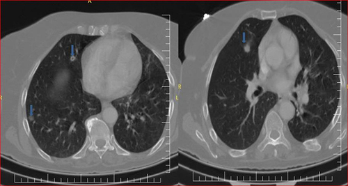
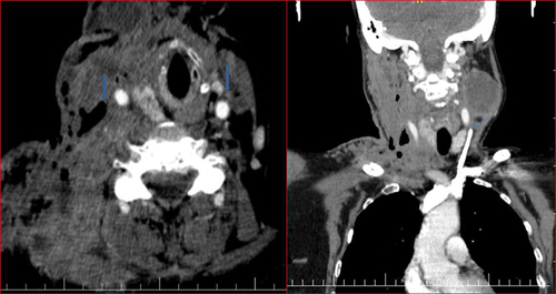
Figure 4 A CT scan of the neck reveals no contrast flow and filling defects in both jugular veins, indicating thrombosis.
Data Sharing Statement
We declared that we had full access to all of the data in this study, and we take complete responsibility for the integrity of the data. All original data are available in the Mogadishu Somali Turkish Training and Research Hospital, Mogadishu, Somalia. Data used to support the findings of this study are available from the corresponding author upon request.