Figures & data
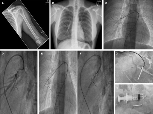
Figure 1 (A) AP radiograph (X-ray) of the left upper arm (humerus and cubital fossa) with no evidence of visible radiopaque subcutaneous contraceptive implant. (B) AP radiograph (X-ray) of the chest. This was initially reported as normal, but closer assessment of the right mid-zone shows the radiopaque contraceptive implant which was initially incorrectly interpreted as a prominent bronchovascular marking given its relatively short, linear outline (black arrow). On subsequent appreciation by the radiologists of the short, tubular shape of the Nexplanon® implant, the foreign body was immediately identified. (C) Judkins right (JR4) catheter positioned in the right pulmonary artery with the contraceptive implant visualized on fluoroscopy (black arrow). (D) Selective pulmonary angiography confirms the intravascular placement of the foreign body (black arrow) in a branch of the right lower lobe pulmonary artery. (E, F) AP and lateral fluoroscopy views demonstrating the utilization of a gooseneck snare to extract the foreign body from the pulmonary artery (black arrows). A straight-ended Terumo (Terumo Corporation, Tokyo, Japan) guidewire was utilized for support. (G, H) Explanted device attached to a Judkins diagnostic (JR4) catheter and the device adjacent to a 5 mL syringe to demonstrate size.