Figures & data
Figure 1 Flexed knee: iliotibial band (1), biceps femoris (2), fibular nerve (3), lateral gastrocnemius tendon (4), and sural nerve and ramifications (5).
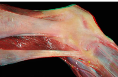
Figure 2 Posterior view of the knee: lateral collateral ligament (1), popliteus tendon (2), popliteofibular ligament (3), and lateral meniscus (4).
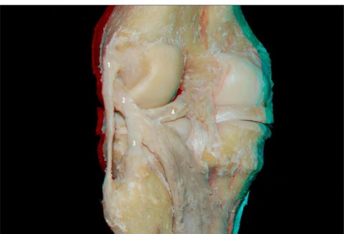
Figure 3 Lateral view of the posterolateral compartment of the extended knee: lateral meniscus (1), lateral collateral ligament (2), popliteus tendon (3), and popliteofibular ligament (4).
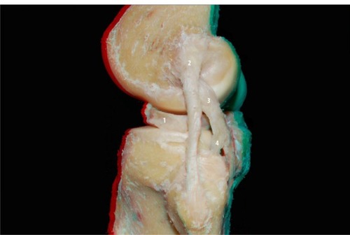
Figure 4 Lateral view of the posterolateral compartment of the flexed knee: lateral collateral ligament (1), popliteus tendon (2), popliteofibular ligament (3), and lateral meniscus (4).
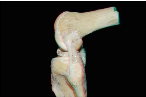
Figure 5 Posterior view of the knee: popliteal artery (1) and ramifications: medial superior genicular artery (2), medial inferior genicular artery (3), lateral superior genicular artery (4), middle genicular artery (5), and lateral inferior genicular artery (6).
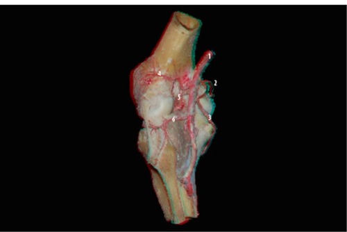
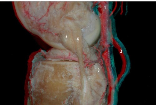
Figure 6 Lateral view of the knee: popliteal artery (1), popliteofibular ligament (2), lateral meniscus (3), lateral inferior genicular artery (4), and lateral superior genicular artery (5).
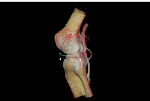
Figure 7 Posterior view of the knee: lateral inferior genicular artery and the posterolateral compartment structures: popliteal artery (1), lateral superior genicular artery (2), medial superior genicular artery (3), posterior septum of the knee (4), lateral inferior genicular artery (5), collateral lateral ligament (6), popliteofibular ligament (7), popliteal muscle (8), and medial inferior genicular artery (9).
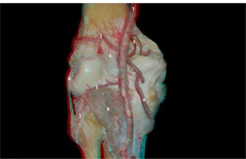
Figure 8 Oblique view of the posterolateral compartment of the knee: the pathway of the lateral inferior genicular artery (5) and the relationship with other structures: lateral superior genicular artery (1), popliteus tendon (2), lateral collateral ligament (3), lateral meniscus (4), and fibular nerve (6).
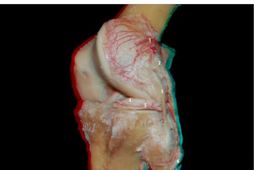
Figure 9 Lateral view of the posterolateral compartment of the knee: the pathway of the lateral inferior genicular artery and the relationship with other structures of the posterolateral compartment: popliteofibular ligament (3), lateral collateral ligament (4), and popliteus tendon (5). Popliteal artery (1) and inferior medial genicular artery (2).