Figures & data
Figure 1 Plantar fascia on the right symptomatic side with a 4mm anterior-posterior diameter as slightly enlarged anterior posterior diameter is usually associated with plantar fasciitis, in this case intra-fascial bleeding (18MHz Matrix linear probe, Canon Medical Systems).
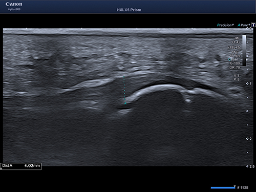
Figure 2 Weight-bearing cone beam CT (WBCT, SCS Med Series H22, 748mGy x cm2) showing a non-healed epiphyseal fracture of the calcaneus eight weeks after a landing in Parkour.
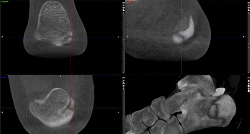
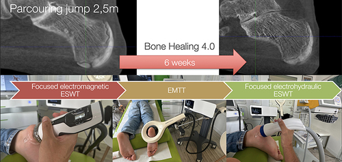
Figure 3 A single bone stimulation session started with focused electromagnetic ESWT, followed by EMTT, leading to focused electrohydraulic ESWT.

Figure 4 Follow-up weight-bearing CT (WBCT) six weeks after the first combined bone stimulation treatment, which was two weeks after the last session with substantial improvement of the delayed union situation at that early point in time.
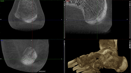
Figure 5 Scheme of a fractioned, repetitive bone stimulation program starting with focused electromagnetic ESWT, followed by EMTT, then thirdly, electrohydraulic focused ESWT in a single session five times in adolescent delayed calcaneus union. Notably, focused ESWT and EMTT did not result in early closure of the calcaneal epiphysis.