Figures & data
Figure 1 MRI findings at onset of neurological disturbances.
Notes: Brain MRI images from June 2015 showed (A) T2 hyperintense lesions in left temporal lobe and brainstem (circles), (B) T1 hyperintense lesions in basal ganglia (thin arrows), known as calcifications, and (C) leptomeningeal contrast enhancement at midbrain–pons junction (arrow).
Abbreviation: MRI, magnetic resonance imaging.
Abbreviation: MRI, magnetic resonance imaging.
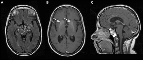
Figure 2 Brain MRI images before and after B-cell depleting therapy.
Notes: Brain MRI images from February 2016 showed extensive and bilateral focal or confluents subcortical and deep WM lesions, hyperintense in long TR sequences, involving (A) semioval centers (S), temporal lobe (*), (B) external capsule, claustrum and subinsular regions (thin arrow) and (C) midbrain (arrow) without contrast enhancement. Moderate subcortical atrophy with dilation of lateral ventricles. Brain MRI in September 2016, 6 months after RTX treatment, showed reductions in numbers and size of hyperintensity lesions in WM, especially in temporal lobes bilaterally (D and E) and brainstem (F).
Abbreviations: MRI, magnetic resonance imaging; WM, white matter; RTX, rituximab.
Abbreviations: MRI, magnetic resonance imaging; WM, white matter; RTX, rituximab.
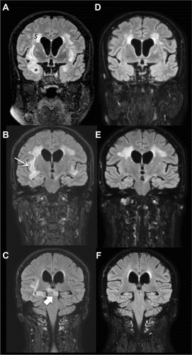
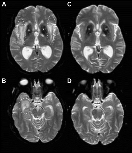
Figure 3 DWI images before and after B-cell depleting therapy.
Notes: Brain MRI DWI sequences from February 2016 (A and B) showed hyperintensity in right temporal lobe (dashed circles), external capsule and claustrum (dashed ovals) with increased apparent diffusion coefficient (not shown) suggesting vasogenic edema. *Basal ganglia calcification. On September 2016, 6 months after RTX treatment, brain MRI DWI sequences (C and D) showed complete disappearance of edema.
Abbreviations: MRI, magnetic resonance imaging; DWI, diffusion-weighted imaging; RTX, rituximab.
Abbreviations: MRI, magnetic resonance imaging; DWI, diffusion-weighted imaging; RTX, rituximab.