Figures & data
Figure 1 Ocular conjunctival findings and skin lesions before the introduction of ustekinumab. Bilateral conjunctival congestion (A) and punctate erythema with desquamation in the forearm (B) and abdomen (C).
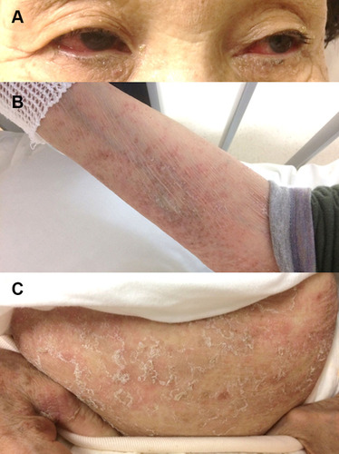
Figure 2 MRI findings of the sacroiliac joints before and two months after the introduction of ustekinumab. (A) STIR-MRI image presents hyperintense foci in the bilateral sacroiliac joints, especially on the right side (arrows). (B) STIR-MRI image shows mild enhancement on the right side only (arrows). MRI-STIR, Short tau inversion recovery-magnetic resonance imaging.
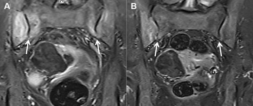
Figure 3 Complete resolution in the bilateral ocular conjunctiva (A), forearm (B), and abdomen (C) two months after one ustekinumab injection.
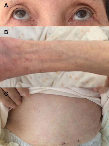
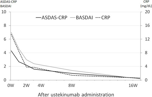
Figure 4 ASDAS-CRP score, BASDAI, and CRP levels after treatment with ustekinumab.