
Figures & data
Figure 1 Liver biopsy is histologically unremarkable on H&E stain (A). On higher magnification, there is intra-hepatocyte brown pigments which represents iron accumulation (B).The pigments appear blue with Perls stain (C). Grade 4 hepatocyte iron overload. The biopsy is negative for significant fibrosis.
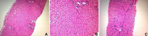
Table 1 Summary of Laboratory Results for Both Patients
Figure 2 Axial T2 weighted image of the liver showed dark signals of the liver (L) relative to normal signals of spleen (S) due to iron deposition.
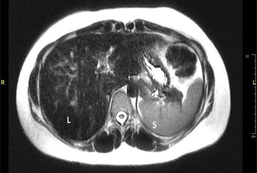
Table 2 Main International Recommendations on Venesection Therapy in HFE Haemochromatosis (with Permission from Powell,2016)Citation34,Citation38,Citation39,Citation40