
Figures & data
Figure 1 Sacroiliitis on MRI. Coronal STIR (short tau inversion recovery) sequence shows extensive subchondral oedema involving the left sacroiliac joint, consistent with unilateral sacroiliitis in a patient with psoriasis.
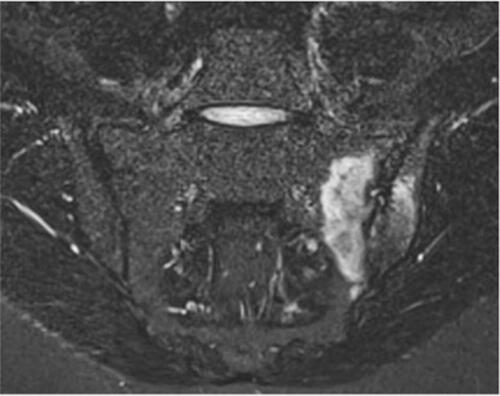
Figure 2 Sacroiliitis on CT. Axial CT (computer tomography; left side of the figure) and T1-weighted sequence (right side of the figure) demonstrates subchondral sclerosis and erosions.
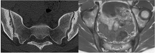
Figure 3 Aseptic spondylodiscitis. Sagittal T1-weighted and STIR sequences in a patient with longstanding psoriatic arthritis show disc oedema with subchondral bone marrow involvement at L2-L3 level, consistent with aseptic spondylodiscitis (Andersson lesion).

Table 1 Imaging Techniques in Psoriatic Arthritis