
Figures & data
Figure 1 The efficacy of aflibercept for treating AMD resistant to ranibizumab (patient 3 with occult CNV).
Notes: A fundus photograph at baseline (A) and SD-OCT images prior to the initial IVR injection (B), at baseline (C), 1 month after the initial IVA injection (D), 3 months after the initial IVA injection (E), and 12 months after the initial IVA injection (F) in a 66-year-old man with occult CNV are shown. Marked SRF and a slight PED have improved at 1-month follow-up examination, and the retinal structure has been maintained over 12 months.
Abbreviations: AMD, age-related macular degeneration; CNV, choroidal neovascularization; IVA, intravitreal aflibercept; IVR, intravitreal ranibizumab; PED, pigment epithelial detachment; SD-OCT, spectral domain optical coherence tomography; SRF, subretinal fluid.
Abbreviations: AMD, age-related macular degeneration; CNV, choroidal neovascularization; IVA, intravitreal aflibercept; IVR, intravitreal ranibizumab; PED, pigment epithelial detachment; SD-OCT, spectral domain optical coherence tomography; SRF, subretinal fluid.
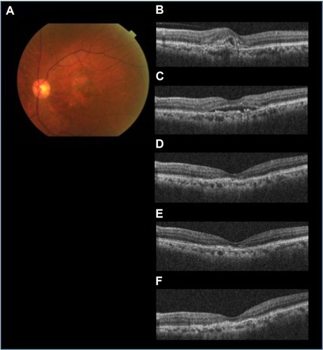
Figure 2 The efficacy of aflibercept for AMD refractory to ranibizumab (patient 8 with PCV).
Notes: A fundus photograph at baseline (A) and SD-OCT images prior to the initial IVR injection (B), at baseline (C), 1 month after the initial IVA injection (D), 3 months after the initial IVA injection (E), and 12 months after the initial IVA injection (F) in a 72-year-old man with PCV are shown. Slight SRF and prominent subfoveal PED have resolved completely at 1-month follow-up examination, and the improved anatomic condition has been maintained over 12 months.
Abbreviations: AMD, age-related macular degeneration; IVA, intravitreal aflibercept; IVR, intravitreal ranibizumab; PCV, polypoidal choroidal vasculopathy; PED, pigment epithelial detachment; SD-OCT, spectral domain optical coherence tomography; SRF, subretinal fluid.
Abbreviations: AMD, age-related macular degeneration; IVA, intravitreal aflibercept; IVR, intravitreal ranibizumab; PCV, polypoidal choroidal vasculopathy; PED, pigment epithelial detachment; SD-OCT, spectral domain optical coherence tomography; SRF, subretinal fluid.
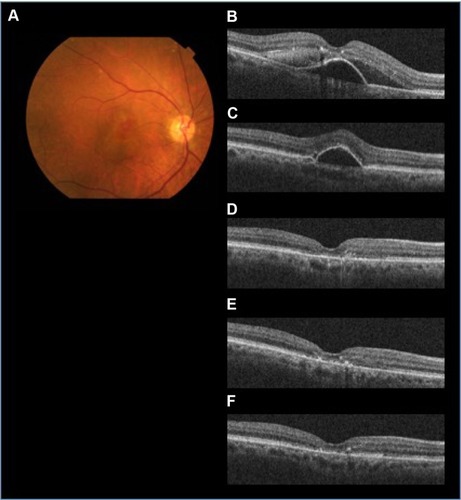
Table 1 Demographics of 14 patients with AMD
Figure 3 The efficacy of aflibercept assessed based on the visual acuity and central macular thickness in 14 patients with AMD (seven patients with occult CNV and seven subjects with PCV).
Notes: The efficacy of aflibercept is assessed based on visual acuity converted to logMAR (A) and the central macular thickness measured by SD-OCT (B). Data obtained at 1 month (1 M), 3 months (3 M), 6 months (6 M), and 12 months (12 M) after the first IVA injection are compared with baseline data. Subcategorical data based on the subtype classifications are also demonstrated (seven patients with occult CNV and seven subjects with PCV). The line charts of the mean data for occult CNV, PCV, and the total cohort are shown in different colors. Statistical significance was defined as P<0.05. Asterisks indicate significant differences (Wilcoxon signed-rank test, *=occult CNV, **=PCV, ***=total cohort).
Abbreviations: AMD, age-related macular degeneration; CNV, choroidal neovascularization; IVA, intravitreal aflibercept; logMAR, logarithm of minimum angle of resolution; PCV, polypoidal choroidal vasculopathy; SD-OCT, spectral domain optical coherence tomography.
Abbreviations: AMD, age-related macular degeneration; CNV, choroidal neovascularization; IVA, intravitreal aflibercept; logMAR, logarithm of minimum angle of resolution; PCV, polypoidal choroidal vasculopathy; SD-OCT, spectral domain optical coherence tomography.
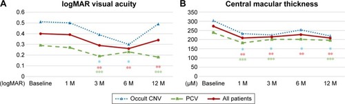
Table 2 Best-corrected visual acuity of 14 patients treated with IVA injection
Table 3 Central macular thickness of 14 patients treated with IVA
Table 4 Anatomical response to IVA treatment in 14 patients with AMD