Figures & data
Figure 1 Schema of PDT.
Notes: (A) The PDT laser spot sometimes cannot cover a large lesion or a peripapillary lesion. (B) Using Ironing PDT, the PDT laser spot is moved at a constant speed, for 83 seconds, to cover the entire lesion. Double-headed arrows show the movement of laser spot.
Abbreviations: AMD, age-related macular degeneration; PDT, photodynamic therapy.
Abbreviations: AMD, age-related macular degeneration; PDT, photodynamic therapy.
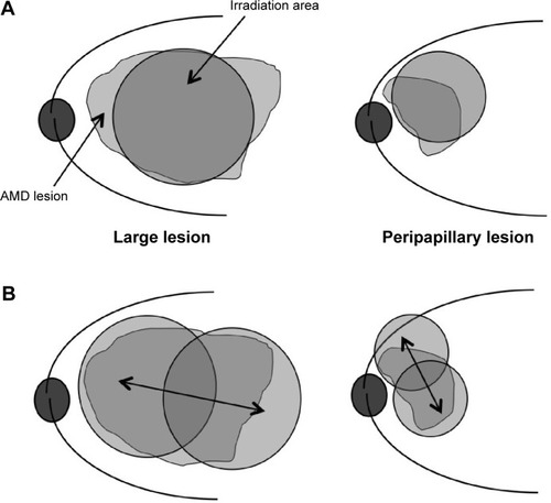
Table 1 Age-related macular degeneration patients treated with Ironing PDT
Figure 2 The change in BCVA (evaluated in logMAR).
Note: The mean logMAR BCVA before treatment was 0.95, and after treatment was 1.09, with no significant difference.
Abbreviations: BCVA, best-corrected visual acuity; logMAR, logarithm of the minimum angle of resolution; PDT, photodynamic therapy.
Abbreviations: BCVA, best-corrected visual acuity; logMAR, logarithm of the minimum angle of resolution; PDT, photodynamic therapy.
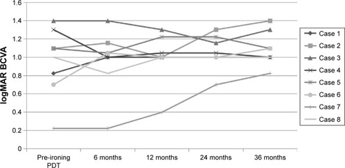
Figure 3 Fundus photography, FA, ICGA, and OCT performed in patient 7.
Notes: (A) Baseline fundus examination shows large serous PED. (B) Late-phase FA shows typical findings of occult CNV and pooling, which demonstrates the presence of serous PED. (C) Baseline OCT shows the presence of type 1 CNV (arrowhead) and serous PED with SRD (white arrow). Inset shows the position of the scanned line (black arrow) of the OCT. (D) After five treatments of IVR, serous PED and SRD still existed, and CNV moved into the serous PED. (E) Late-phase FA shows strong leakage into the PED, indicative of fibrovascular PED. (F) OCT shows a hyper-reflective lesion under the RPE, with SRD (white arrows). Inset shows the position of the scanned line (black arrow) of the OCT.
Abbreviations: CNV, choroidal neovascularization; FA, fluorescein angiography; ICGA, indocyanine green angiography; IVR, intravitreal injections of ranibizumab; OCT, optical coherence tomography; PED, pigment epithelial detachment; RPE, retinal pigment epithelium; SRD, serous retinal detachment.
Abbreviations: CNV, choroidal neovascularization; FA, fluorescein angiography; ICGA, indocyanine green angiography; IVR, intravitreal injections of ranibizumab; OCT, optical coherence tomography; PED, pigment epithelial detachment; RPE, retinal pigment epithelium; SRD, serous retinal detachment.
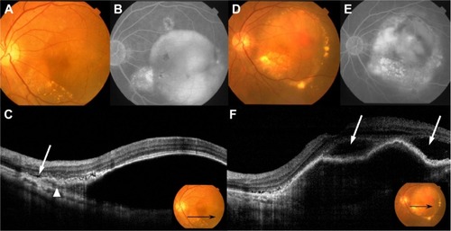
Figure 4 Ironing PDT performed in patient 7.
Notes: (A) Because the mean GLD was 9,980 μm, conventional PDT could not cover the entire lesion. (B) The laser spot was moved at a constant speed, for 83 seconds, to cover the entire lesion using a 7,200 μm laser spot (dashed line), after a 1,000 μm margin was added to the smaller diameter of the lesion. Double-headed arrow shows the movement of laser spot.
Abbreviations: GLD, greatest linear dimension; PDT, photodynamic therapy.
Abbreviations: GLD, greatest linear dimension; PDT, photodynamic therapy.
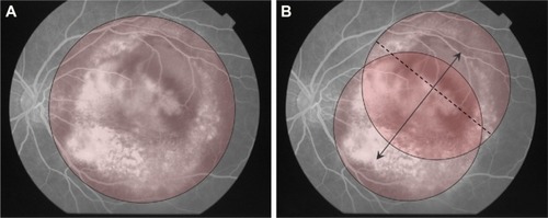
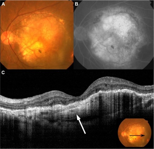
Figure 5 Fundus photography, FA and OCT 36 months after the first Ironing PDT performed in patient 7.
Notes: (A) Fundus examination shows CNV was scarred and exudation was resolved. (B) Late-phase FA shows hyperfluorescence of fibrosis. (C) OCT shows a hyperreflective lesion under the RPE (white arrow), but exudation was not observed. Inset shows the position of the scanned line (black arrow) of the OCT.
Abbreviations: CNV, choroidal neovascularization; FA, fluorescein angiography; OCT, optical coherence tomography; PDT, photodynamic therapy; RPE, retinal pigment epithelium.
Abbreviations: CNV, choroidal neovascularization; FA, fluorescein angiography; OCT, optical coherence tomography; PDT, photodynamic therapy; RPE, retinal pigment epithelium.