Figures & data
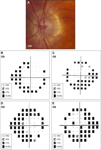
Figure 1 Patient 1.
Notes: (A) OD optic disk image shows vertically oval cup with INF thinning of the NRR. (B) OS optic disk image with media haze and diffuse disk pallor. (C) OD Goldmann perimetry with diffuse constriction, SUP > INF. (D) OS Goldmann perimetry shows remaining island of central and inferotemporal visual field. (E) Stratus OCT RNFL thickness analysis OD shows no RNFL loss. (F) OS RNFL quadrant and analysis show severe losses (<1% of normative data) in the NAS and TEMP quadrants. OS RNFL clock hour analysis corroborates RNFL thickness findings. OD RNFL quadrant and clock hour analyses were both normal.
Abbreviations: INF, inferior; NRR, neuroretinal rim; SUP, superior; OCT, optical coherence tomography; RNFL, retinal nerve fiber layer; NAS, nasal; TEMP, temporal; NA, not applicable; S, superior; N, nasal; T, temporal; I, inferior.
Abbreviations: INF, inferior; NRR, neuroretinal rim; SUP, superior; OCT, optical coherence tomography; RNFL, retinal nerve fiber layer; NAS, nasal; TEMP, temporal; NA, not applicable; S, superior; N, nasal; T, temporal; I, inferior.
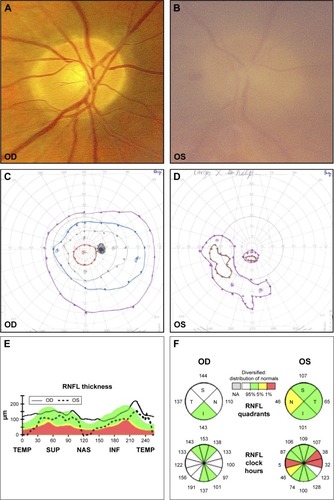
Figure 2 Patient 2.
Notes: (A) OD disk image showing fibrous membrane over disk with inferior thinning of NRR. There is no NVD. (B) OS optic disk appears hyperemic with dense fibrous membrane. There is no NVD. (C) OD Goldmann perimetry shows superior visual field loss. (D) OS Goldmann perimetry shows mild constriction of the superotemporal visual field.
Abbreviations: NRR, neuroretinal rim; NVD, neovascularization of the disk.
Abbreviations: NRR, neuroretinal rim; NVD, neovascularization of the disk.
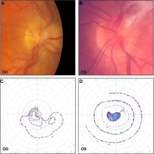
Figure 3 Patient 3.
Notes: (A) OD optic disk has pink and regular NRR without peripapillary atrophy. (B) OS optic disk was similar with fibrous membrane and vessel sheathing. (C) Initial OD Goldmann perimetry with relatively intact visual field. (D) Initial OS Goldman perimetry with superotemporal constriction. (E) Interval OD Goldmann visual field after 14 years shows marked visual constriction with central scotoma (OS unavailable since the patient’s left eye became phthisical). (F) OD stratus OCT shows mild RNFL thinning superiorly, otherwise RNFL thickness is normal. (G) Normal RNFL quadrant and RNFL clock hour analyses OD.
Abbreviations: NRR, neuroretinal rim; OCT, optical coherence tomography; RNFL, retinal nerve fiber layer; TEMP, temporal; SUP, superior; NAS, nasal; INF, inferior; NA, not applicable; S, superior; N, nasal; T, temporal; I, inferior.
Abbreviations: NRR, neuroretinal rim; OCT, optical coherence tomography; RNFL, retinal nerve fiber layer; TEMP, temporal; SUP, superior; NAS, nasal; INF, inferior; NA, not applicable; S, superior; N, nasal; T, temporal; I, inferior.
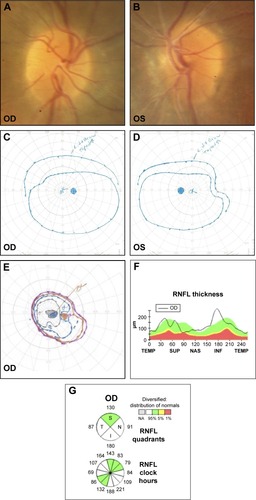
Figure 4 Patient 4.
Notes: (A and B). Bilateral disk pallor with loss of disk capillaries and attenuation of major vessels, suggestive of optic atrophy. (C) Goldmann perimetry with severe visual field loss and preservation of central vision OD. (D) Goldmann perimetry with severe visual field loss and split fixation OS. (E) Stratus OCT RNFL thickness analysis OU showed marked SUP, NAS, and INF thinning (<1% of normal). (F) RNFL quadrant and clock hours were consistent with the RNFL thickness findings.
Abbreviations: OCT, optical coherence tomography; RNFL, retinal nerve fiber layer; SUP, superior; NAS, nasal; INF, inferior; TEMP, temporal; NA, not applicable; S, superior; N, nasal; T, temporal; I, inferior.
Abbreviations: OCT, optical coherence tomography; RNFL, retinal nerve fiber layer; SUP, superior; NAS, nasal; INF, inferior; TEMP, temporal; NA, not applicable; S, superior; N, nasal; T, temporal; I, inferior.
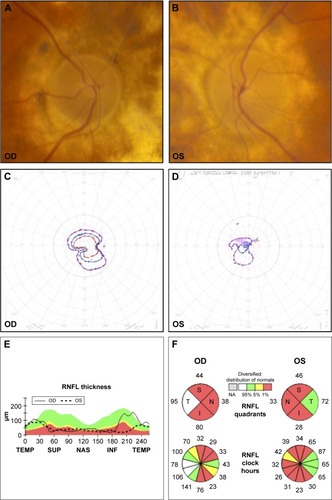
Figure 5 Patient 5.
Notes: (A) OS optic disk has pink and regular NRR (OD examination was similar to OS examination). (B and C) Humphrey Visual Field 24-2 SITA-Standard protocol with superior and inferior arcuate defects OS > OD, and superior and inferior nasal step defects OU. (D and E) Interval Humphrey Visual Field 24-2 SITA-Standard protocol (obtained 6 years later) demonstrates glaucomatous progression with denser superior arcuate defects OU.
Abbreviation: NRR, neuroretinal rim.
Abbreviation: NRR, neuroretinal rim.