Figures & data
Figure 1 Upper eyelid interferes with the visual axis causing stimulus deprivation or induces amblyogenic astigmatism.
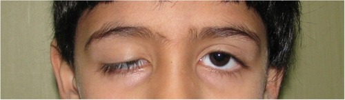
Table 1 Choice of surgical technique according to the grade of ptosis and levator function
Figure 2 Fox pentagon technique.
Notes: (A) Skin mark. (B) Wright’s fascial needle. (C) Control the eyelid shape. (D) Nylon tied together.
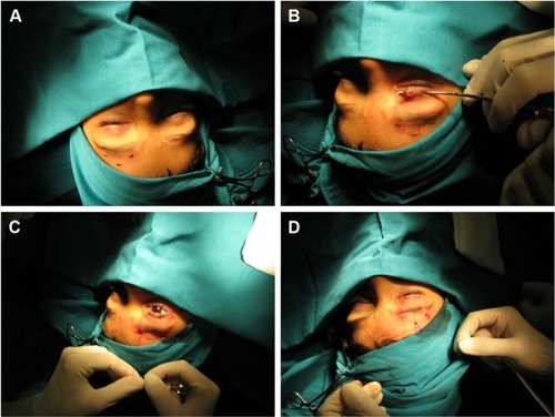
Figure 3 (A) Preoperative and (B) postoperative result of frontalis sling procedure in a case of severe ptosis.
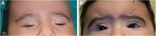
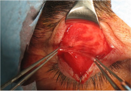
Figure 4 Aponeurosis of the levator muscle.