
Figures & data
Figure 1 Protrusion of orbital contents.
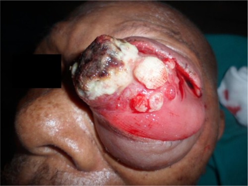
Figure 2 CT scan of the orbits.
Note: Intraorbital tumor without orbital bony destruction.
Abbreviation: CT, computed tomography.
Abbreviation: CT, computed tomography.
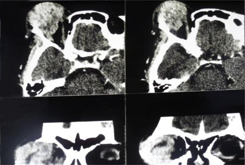
Figure 3 Histological examination (magnification ×40).
Notes: (A) Squamous cell carcinoma (the white arrows indicate nodula with concentric laminated layer called “keratinous pearls”). (B) Adenoid cystic carcinoma (the white arrows indicate cribriform mass of dyscaryotic tumor cells). (C) Achromic melanoma (the white arrows indicate Proliferative pearls of achromic melanocytes).

Figure 4 Spontaneous healing of the orbital cavity.
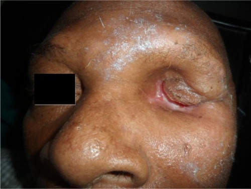
Table 1 Baseline demographic and clinical characteristics of patients included in this study