
Figures & data
Figure 1 Reperfusion in eye of a 61-year-old male, following treatment with 5 injections of ranibizumab.
Notes: Preinjection fluorescein angiography (A–C) shows ischemia and abnormal neovascularization (A, preinjection fluorescein angiogram; B, magnified view of preinjection fluorescein angiogram; and C, binary preinjection image). Postinjection images (D–F) show reperfusion with improved perfusion as compared with preinjection image A (D, postinjection fluorescein angiogram; E, magnified view of preinjection fluorescein angiogram; and F, binary postinjection image).
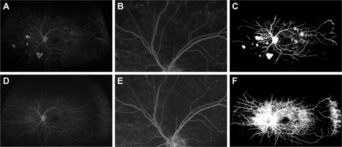
Figure 2 Reperfusion in eye of a 60-year-old female, following treatment with two injections of ranibizumab.
Notes: The preinjection fluorescein angiography (A, preinjection fluorescein angiogram and B, binary preinjection image) shows peripheral ischemia both inside and outside the field of 7SF FA (B, red outline). The postinjection image (C, postinjection fluorescein angiogram and D, binary postinjection image) shows reperfusion both inside and outside the field of 7SF FA (D, red outline).
Abbreviations: 7SF, 7-standard field; FA, fluorescein angiography.
Abbreviations: 7SF, 7-standard field; FA, fluorescein angiography.
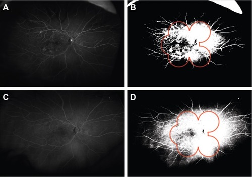
Table 1 Patient demographics (n=15 patients)
Table 2 Treatment and UWFA imaging (n=16 eyes)