
Figures & data
Figure 1 Clinical appearance of a patient with Coats’ disease. (A and B) The fundus image of a patient with Coats’ disease with exudative total retinal detachment. There is central arteriolar mild dilation and tortuosity. Peripheral telangiectatic vessels along aneurysmal changes are visible. Many microvascular shunts nearly in 360° of periphery and midperiphery of retina are evident. (C and D) Angiographic finding as telangiectatic vessels, microaneurysms, sacular aneurysms, shunt vessels and peripheral avascular area in temporal and inferior retina.
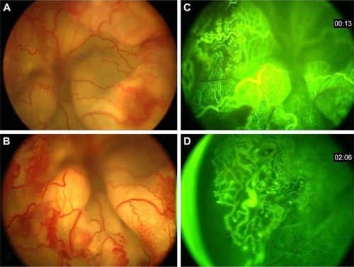
Table 1 Coats’ disease based on the age category: demographic features
Table 2 Coats’ disease based on the age category: comparison of serum values with normal controls
Table 3 Coats’ disease comparison with controls: adjusted to age and sex as confounding factors (logistic regression)