Figures & data
Figure 1 MRI of a patient with posterior uveal melanoma: arrow + red color indicate tumor mass, yellow and blue colors indicate lens, and green and orange colors indicate optic nerves.
Abbreviation: MRI, magnetic resonance imaging.
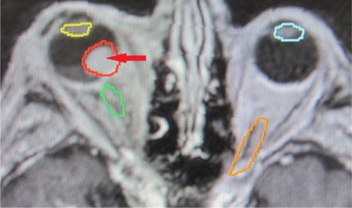
Figure 2 Stereotactic planning scheme of the same patient.
Note: Arrow and red circle indicate tumor mass.
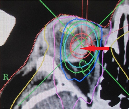
Figure 3 Virtual model of the eye, outer view; arrow indicates optic nerve.
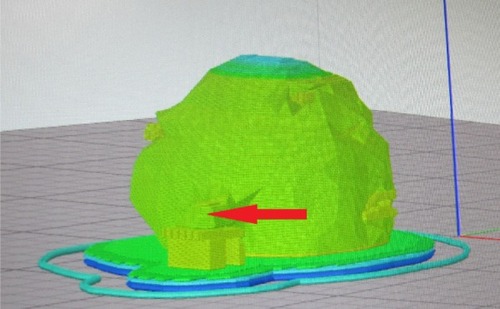
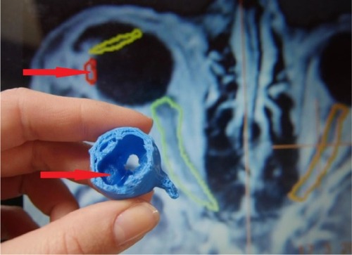
Figure 4 Printed 3D model of the eye with intraocular uveal melanoma, middle-stage T2; arrow indicates tumor mass.
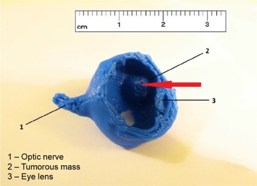
Figure 5 Printed 3D model of the eye with intraocular uveal melanoma, middle-stage T2 (arrows indicate tumor mass), with stereotactic planning scheme on the background.