
Figures & data
Table 1 Prevalence of IPCV among 129 patients with angiographic CNVs
Figure 1 Fundus findings of most patients in the study.
Notes: Red arrows represent subretinal hemorrhage and white arrows represents subretinal lipid exudate. Drusens are noted in the peripheral fundus (yellow arrows).
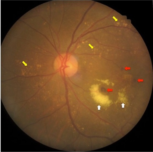
Figure 2 Indocyanine green angiographic findings of IPCV lesions: polypoidal lesions with hypofluorescent halos (red arrows) and branching vascular networks (area within yellow dotted line).
Abbreviation: IPCV, idiopathic polypoidal choroidal vasculopathy.
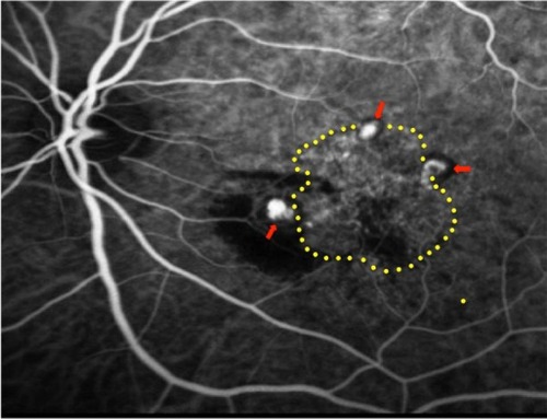
Table 2 The characteristics of patients with IPCV in the study
Table 3 The characteristics of IPCV patients in this study compared to the previous studies