Figures & data
Figure 1 Case 1: (A) Endoscopic photograph of the right nasal cavity showing the palatal defect, exposed bone of the lateral wall in the area of maxillary sinus and altered anatomy of the lateral wall. (B) The 3D CT-DCG showed absence of the right maxilla with right dilated lacrimal sac and an abrupt obstruction at the sac–duct junction. The DCG findings of the left lacrimal apparatus were normal.
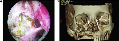
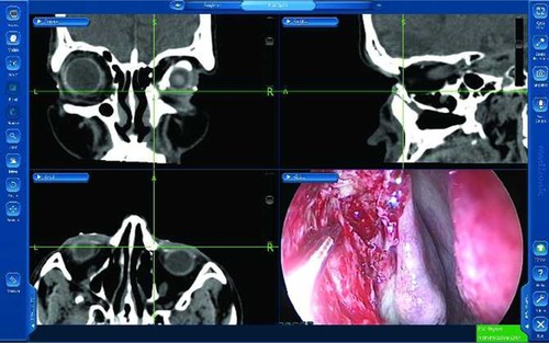
Figure 2 Case 1: Image-guided dacryolocalization view showing the coronal (upper left), sagittal (upper right) and axial (lower left) CT images, as well as the endoscopic view (lower right).
Abbreviation: CT, computed tomography.
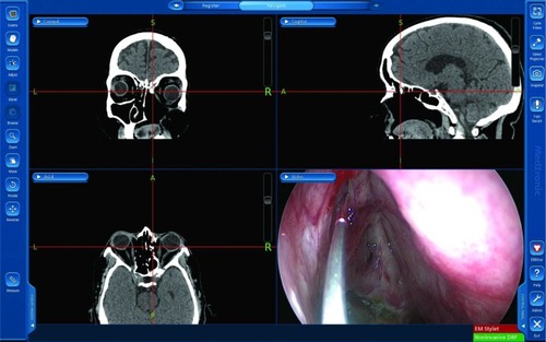
Figure 3 Case 1: Intraoperative image-guided view depicting the image-guided dacryolocalization using the 3D-reconstructed virtual model of the DCG (lower right panel).
Abbreviation: DCG, dacryocystography.
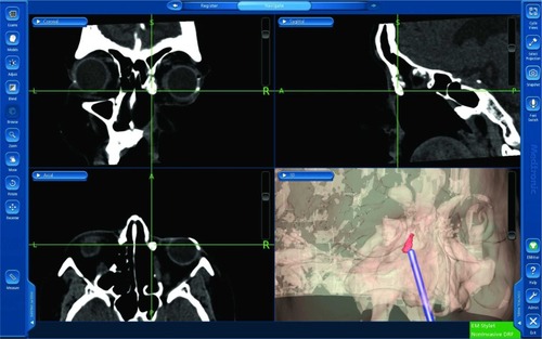
Figure 4 Case 2: (A) Endoscopic view of the left nasal cavity showing alteration of the nasal anatomy. (B) The 3D CT-DCG shows an irregular dilated left lacrimal sac and an abrupt obstruction at the level of the proximal nasolacrimal duct. The DCG findings of the right lacrimal apparatus were normal.
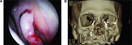
Figure 5 Case 2: (A) Image-guided dacryolocalization view showing the CT images and the endoscopic view (lower right). Note that the intersection of crosshairs shows the simultaneous endoscopic localization of the contrast-filled lacrimal sac. (B) Active endoscopic real-time tracking of the lacrimal apparatus using the 3D virtual model.
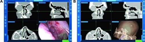
Figure 6 Case 3: (A) The 3D CT-DCG showed a regularly dilated right lacrimal sac and an abrupt obstruction at the level of the sac–duct junction. The DCG findings of the left lacrimal apparatus showed multiple areas of stenosis at the sac–duct junction and within the nasolacrimal duct. (B) Endoscopic localization of the contrast-filled sac. Note that the green crosshairs depict the accurate localization on all the CT images.
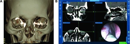
Figure 7 Case 3: Intraoperative active localization of the lacrimal apparatus following the osteotomy.