Figures & data
Figure 1 Subpterygium injection of MMC and bevacizumab by insulin syringe under topical anesthesia.
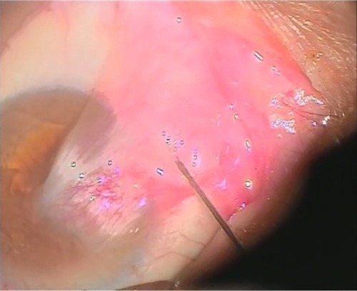
Table 1 Preoperative data of 20 patients involved in our study
Table 2 The data of the two cases used as control
Figure 2 Grades of recurrence.
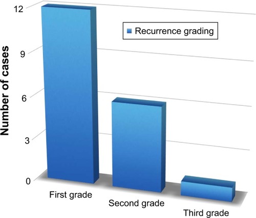
Figure 3 A photomicrograph of untreated pterygium section showing alternative thin and thick areas that appear in the surface epithelium.
Abbreviation: H&E, haematoxilin and eosin stain.
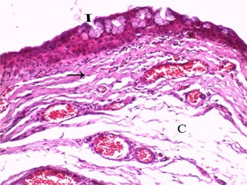
Figure 4 A photomicrograph of untreated pterygium section showing connective tissue stroma that is covered by conjunctival epithelium.
Abbreviation: H&E, haematoxilin and eosin stain.
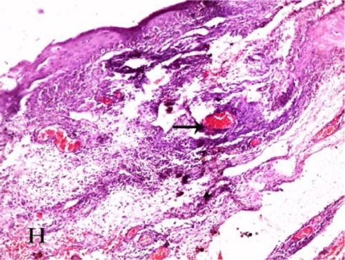
Figure 5 A photomicrograph of untreated pterygium section showing subepithelial connective tissue that is rich in collagen fibers.
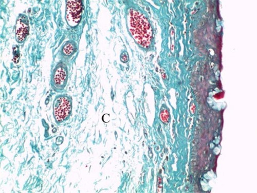
Figure 6 A photomicrograph of noninjected pterygium section immunostained with CD31 antibody showing numerous CD31-positive vessels in subepithelial and connective tissue stroma.
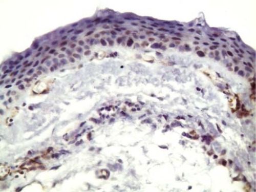
Figure 7 A photomicrograph of treated pterygium section showing the absence of goblet cells in the covering conjunctival epithelium.
Abbreviation: H&E, haematoxilin and eosin stain.
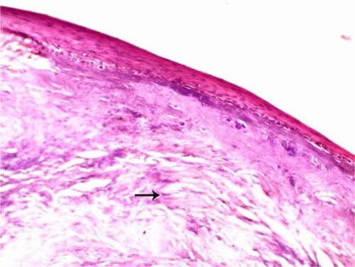
Figure 8 A photomicrograph of treated pterygium section showing the collagen fibers that are regularly arranged close to each other with no signs of degeneration.
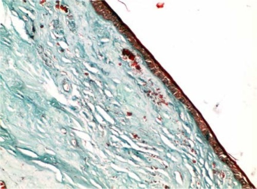
Figure 9 A photomicrograph of injected pterygium-immunostained section showing a small number of subepithelial CD31-positive vessels.
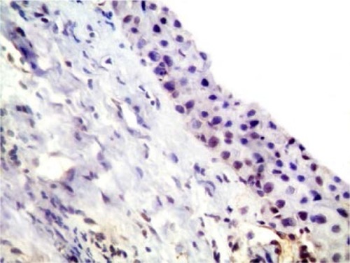
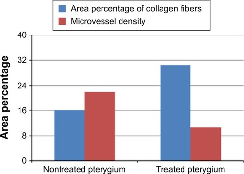
Figure 10 The mean of area percentage of collagen fibers and microvessel density of CD31 of the two groups.