Figures & data
Figure 1 Slit lamp photograph of the type 1 Boston Keratoprosthesis with epithelial growth over the optic.
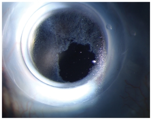
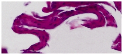
Figure 2 Hematoxylin and eosin stain of epithelial growth removed from the type 1 Boston Keratoprosthesis optic surface showing a variable thickness and number of cell layers, no vascular structures, no goblet cells, and no basement membrane.