Figures & data
Figure 1 Left eye of 12-year-old male.
Notes: Fundus photograph showing presence of hard exudates and arteriolar aneurysms on the disk and along the superotemporal arcades (A); fluorescein angiography in the early arteriovenous phase (B) showing aneurysmal abnormalities that leak, along with staining of the optic disk suggestive of neuroretinitis and focal staining of the veins suggestive of vasculitis (C).
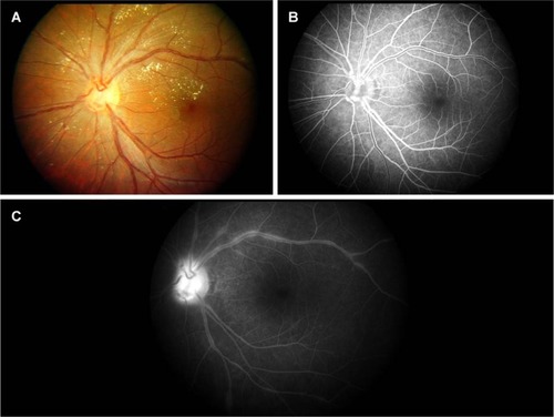
Figure 2 Fundus photography of eyes of 12-year-old male.
Notes: Hard exudates and arteriolar aneurysms along the superotemporal and inferotemporal arcades (A); left eye, showing presence of hard exudates and arteriolar aneurysms on the disk and along the superotemporal arcades (B); after treatment with laser photocoagulation 4 years later, there was a decrease in hard exudates and in arteriolar aneurysms in both eyes (C, D).
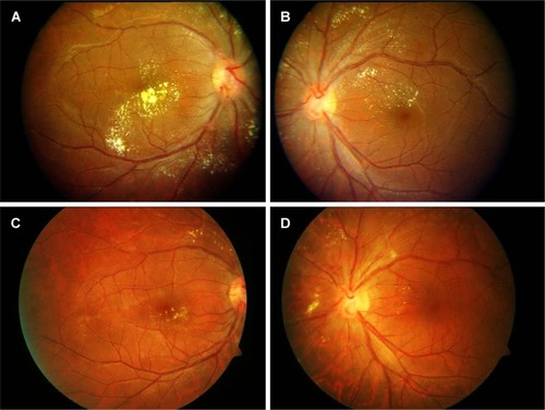
Figure 3 Left eye of an 11-year-old girl who presented with mild blurring of vision.
Notes: Fundus photograph showing deposition of hard exudates in the posterior pole, as well as in the peripapillary location (A); fluorescein angiography of the left eye in the early lamellar venous phase, showing the hyperfluorescent knob-like aneurysmal dilatations giving the classic knot-like appearance of the arterioles (B), along with diffuse leakage in the late phase (C).
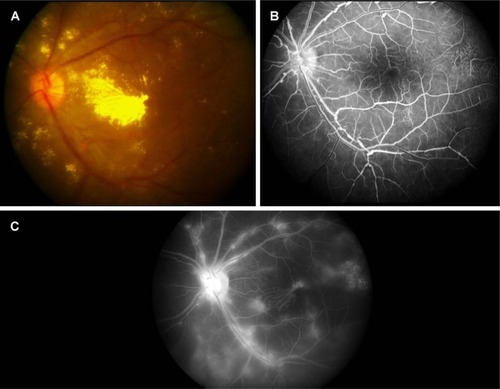
Figure 4 Fundus photography of both eyes of an 11-year-old girl who presented with blurring of vision both eyes.
Notes: Extensive deposition of hard exudates in the posterior pole and peripapillary location, along with aneurysmal dilatation of arteries (A, B); after treatment with laser photocoagulation and multiple dexamethasone injections 7 years later, there was a decrease in hard exudates, along with a decrease in arteriolar aneurysms (C, D).
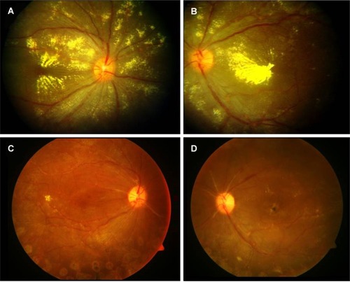
Figure 5 Left eye of an 11-year-old IRVAN patient.
Notes: Fundus photograph showing neovascularization of the optic disk with vitreous hemorrhage (A); fluorescein angiography showing early hyperfluorescence of aneurysms (B) and leakage from neovascularization of disk and aneurysmal dilatations, along with extensive capillary dropout temporally (C).
Abbreviation: IRVAN, idiopathic retinal vasculitis, aneurysms, and neuroretinitis.
Abbreviation: IRVAN, idiopathic retinal vasculitis, aneurysms, and neuroretinitis.
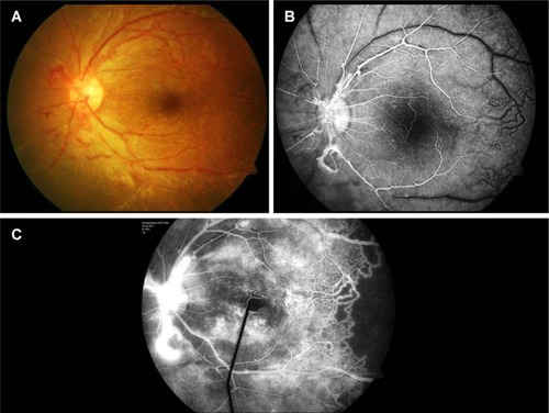
Figure 6 Wide-field fluorescein angiography in a case of IRVAN; an 11-year-old girl who presented with blurring of vision both eyes.
Notes: Early hyperfluorescence of aneurysms, extensive capillary dropout in the periphery, and small neovascularization elsewhere (A); post-treatment, showing panretinal photocoagulation laser scars in the periphery with persistent leakage from aneurysmal dilatations and neovascularization elsewhere, in addition to resolution of upper nasal aneurysms (B).
Abbreviation: IRVAN, idiopathic retinal vasculitis, aneurysms, and neuroretinitis.
Abbreviation: IRVAN, idiopathic retinal vasculitis, aneurysms, and neuroretinitis.
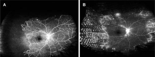
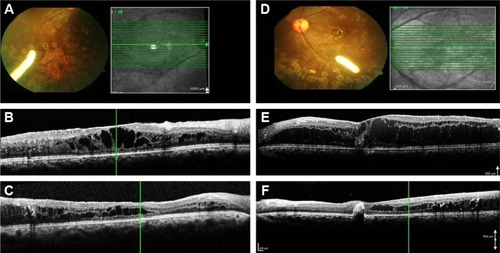
Figure 7 Right and left eye of a case of IRVAN; a 12 years-old-girl who presented with blurring of vision both the eyes.
Notes: Fundus photography (A, D) of an Ozurdex implant in situ, showing decrease in the macular edema on optical coherence tomography after combined treatment with laser and injection (B, C, E, F).
Abbreviation: IRVAN, idiopathic retinal vasculitis, aneurysms, and neuroretinitis.
Abbreviation: IRVAN, idiopathic retinal vasculitis, aneurysms, and neuroretinitis.