
Figures & data
Table 1 IWOS criteria for diagnosing ocular sarcoidosis: introducing seven clinical signs suggestive of ocular sarcoidosis, five laboratory investigations in suspected ocular sarcoidosis, and four levels of certainty
Table 2 Spectrum of ocular manifestations of sarcoidosis
Figure 1 Sarcoid-related panuveitis with vitreous haze and right-eye optic disk swelling.
Note: ×1.84 at 50° (TRC-50DX; Topcon Corporation, Tokyo, Japan).
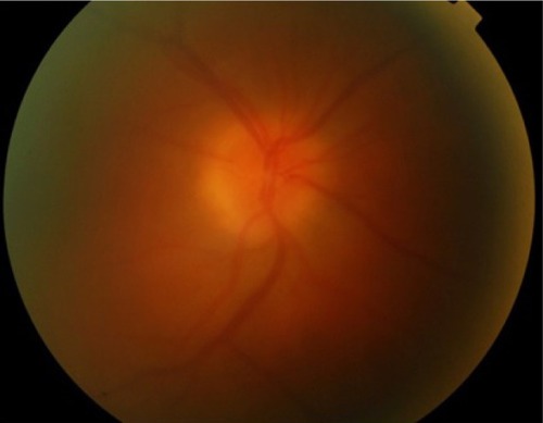
Figure 2 Spectral-domain optical coherence tomography.
Note: Optic nerve-head granuloma, adjacent choroidal neovascular membrane with inner segment/outer segment (ellipsoid zone) disruption, and small amount of subretinal fluid in lung biopsy-proven sarcoidosis patient.
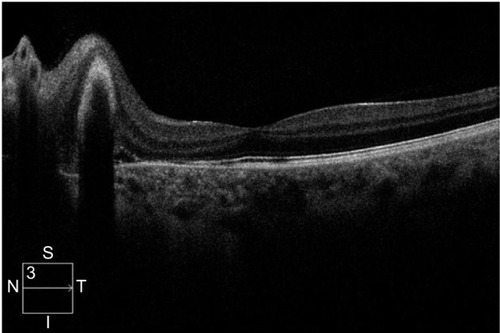
Table 3 Treatment options in sarcoidosis induced uveitis according to location of inflammation and severity
Table 4 Recent studies evaluating the use of adalimumab in sarcoid-associated uveitis