Figures & data
Figure 1 (A) Fundoscopy image of ocular melanoma; (B) measurements of height and maximum base diameter for choroidal melanoma on ultrasound scan.
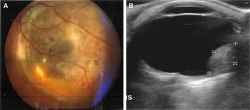
Figure 2 (A) Characteristic homogeneous appearance of choroidal melanoma on ultrasound scans; (B) application of Doppler technique depicting and enabling the study of tumor’s neovascularization.
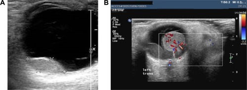
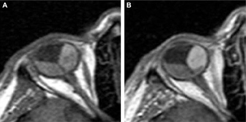
Figure 3 Characteristic appearance of choroidal melanoma on MRI.
Abbreviation: MRI, magnetic resonance imaging.
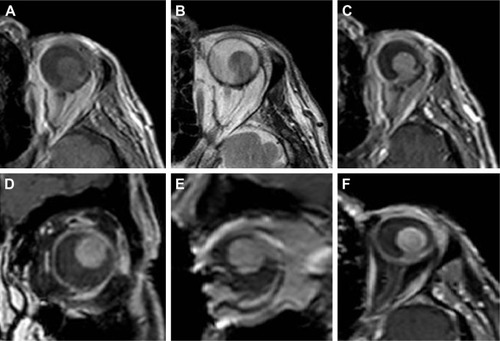
Figure 4 Homogeneity incidence detected by using different methods.
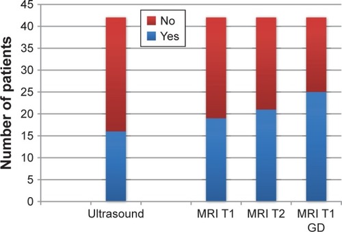
Figure 5 Number of cases where retinal detachment was detected by using different methods.
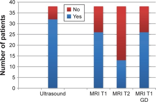
Figure 6 Imaging of choroidal melanoma on MRI and ultrasound scan.
Abbreviation: MRI, magnetic resonance imaging.
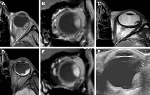
Figure 7 Choroidal melanoma on MRI and ultrasound scan.
Abbreviation: MRI, magnetic resonance imaging.
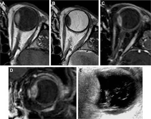
Figure 8 Arterial enhancement of choroidal melanoma on MRI scan enabling imaging differentiation between the tumor and retinal detachment.
Abbreviation: MRI, magnetic resonance imaging.