Figures & data
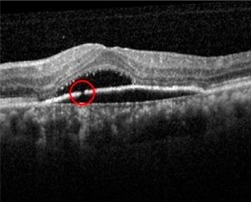
Figure 2 RPE discontinuity and subretinal fluid.
Notes: Area of discontinuity shown in the red circle. This image was originally published in the Retina Image Bank. Avris Romario Diparaja Siahaan. Discontinuity RPE. Retina Image Bank. 2014; Image Number 20572. © the American Society of Retina Specialists.15
Abbreviation: RPE, retinal pigment epithelium.
Abbreviation: RPE, retinal pigment epithelium.

