Figures & data
Table 1 One-year follow-up of intravitreal aflibercept for choroidal neovascularization associated with chorioretinitis
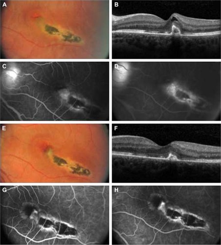
Figure 1 (A) Baseline fundus photographs (OS) show a macular scar with fibrosis, subretinal fluid, and hemorrhage. (B) Baseline optical coherence tomographic (OCT) scan shows increased retinal thickening and a highly reflective subretinal complex. (C and D) Baseline early- and late-phase fluorescein angiographic images show leakage from choroidal neovascularization. (E) Posttreatment fundus photographs taken at 4 months show a macular scar and no evidence of subretinal fluid or hemorrhage. (F) Posttreatment OCT scan taken at 4 months shows decreased retinal thickness and a persistent highly reflective subretinal complex. (G and H) Posttreatment early- and late-phase fluorescein angiographic images taken at 4 months show no evidence of leakage.