
Figures & data
Table 1 Preoperative characteristics of all patients
Figure 1 Anterior segment optical coherence tomography of the interface fluid as optical space between the corneal flap and the stroma.
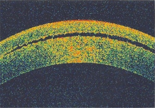
Figure 2 Corneal topography shows thickening of the cornea due to flap edema and interface fluid.
Abbreviations: Thk, thickness; Curv, curvature; Rbf, radial basic function; OS, occulus sinister [left eye].
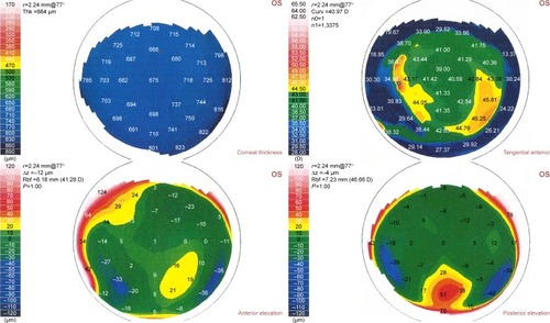
Table 2 Postoperative characteristics of IFS group
Table 3 Comparison between the preoperative characteristics of IFS group and control group (LASIK patients who did not develop IFS)