181
Views
35
CrossRef citations to date
0
Altmetric
Review

Ultra-wide-field fluorescein angiography in diabetic retinopathy: a narrative review

, , , , , , , , & show all
Pages 803-807 | Published online: 27 Apr 2017

Figures & data

Figure 1 Ultra-wide-field fluorescein angiography (UWFA) of a 68-year-old male patient affected by proliferative diabetic retinopathy in both the eyes. Early phases of the right (A) and left (B) eyes. Late phases of the right (C) and left (D) eyes. In early angiographic phases, UWFA discloses hyperfluorescent dots in both the eyes suggestive of microaneurysms and broad peripheral and mid peripheral areas of capillary non-perfusion. In late frames, UWFA discloses peripheral perivascular and mild macular dye leakage, suggestive of blood–retina barrier disruption, and intense hyperfluorescence of retinal surface, indicative of epiretinal neovascularization in both the eyes.

Figure 1 Ultra-wide-field fluorescein angiography (UWFA) of a 68-year-old male patient affected by proliferative diabetic retinopathy in both the eyes. Early phases of the right (A) and left (B) eyes. Late phases of the right (C) and left (D) eyes. In early angiographic phases, UWFA discloses hyperfluorescent dots in both the eyes suggestive of microaneurysms and broad peripheral and mid peripheral areas of capillary non-perfusion. In late frames, UWFA discloses peripheral perivascular and mild macular dye leakage, suggestive of blood–retina barrier disruption, and intense hyperfluorescence of retinal surface, indicative of epiretinal neovascularization in both the eyes.

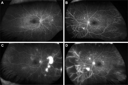
Table 1 Synoptic table on current literature regarding ultra-wide-field fluorescein angiography in diabetic retinopathy