
Figures & data
Table 1 Patient characteristics according to the type of choroidal neovasculopathy
Figure 1 Incidence of ORT.
Notes: (A) Overall incidence of ORT among the included patients. (B) Incidence of ORT among the included patients according to the type of CNV. *P<0.01.
Abbreviations: ORT, outer retinal tubulation; CNV, choroidal neovascularization; t-AMD, typical age-related macular degeneration; PCV, polypoidal choroidal vasculopathy; RAP, retinal angiomatous proliferation; mCNV, myopic choroidal neovascularization.
Abbreviations: ORT, outer retinal tubulation; CNV, choroidal neovascularization; t-AMD, typical age-related macular degeneration; PCV, polypoidal choroidal vasculopathy; RAP, retinal angiomatous proliferation; mCNV, myopic choroidal neovascularization.
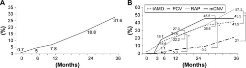
Figure 2 Pathological changes in optical coherence tomography findings that precede the formation of subretinal hyperreflective material and ORT.
Notes: (A) A case of PCV with overlying SHRM. At baseline, a large retinal pigment epithelial detachment, an SHRM (white arrow head), and an abnormal vascular network are observed as a double layer. At 7 months, the boundary of the SHRM becomes clear. At 22 months, two large open ORTs (white arrow head) are seen on either side of the fovea, containing hyperreflective dots inside; the SHRM becomes a heterogeneous mass (white arrow). At 28 months, segmental small ORT (white arrow head) is seen on the homogeneous subretinal fibrosis (white arrow). (B) A case of t-AMD with mixed lesions containing type-1 and -2 CNV components. At baseline, the CNV directly intrudes into the outer layer of the retina, and the RPE line is disrupted (white arrow head). Exudative changes with intra-retinal fluid and fibrin can be seen. At 4 months, the scarring CNV becomes a more solid mass, and the RPE line is disrupted across a wide range. At 15 months, a small ORT (white arrow head) is seen on a heterogeneous SHRM (white arrow). The stump of the remaining ISe line is rolled on the opposite side (asterisk). At 25 months, the hyperreflective mass became homogeneous (white arrow). The ORT cannot be recognized because of the large amount of intraretinal fluid.
Abbreviations: ORT, outer retinal tubulation; PCV, polypoidal choroidal vasculopathy; SHRM, subretinal hyperreflective material; t-AMD, typical age-related macular degeneration; CNV, choroidal neovascularization; RPE, retinal pigment epithelium; VA, visual acuity; Mo, months; ISe, inner segment ellipsoid.
Abbreviations: ORT, outer retinal tubulation; PCV, polypoidal choroidal vasculopathy; SHRM, subretinal hyperreflective material; t-AMD, typical age-related macular degeneration; CNV, choroidal neovascularization; RPE, retinal pigment epithelium; VA, visual acuity; Mo, months; ISe, inner segment ellipsoid.
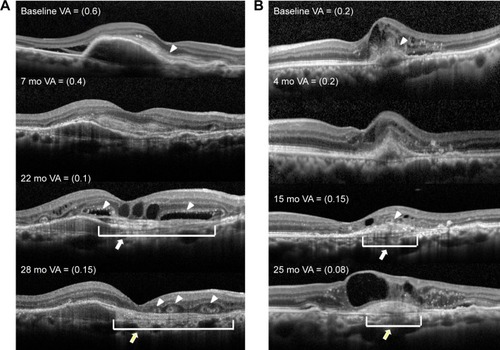
Figure 3 Incidence of subretinal fibrosis.
Notes: (A) Overall incidence of subretinal fibrosis among the included patients. (B) Incidence of subretinal fibrosis among the included patients according to type of CNV.
Abbreviations: CNV, choroidal neovascularization; t-AMD, typical age-related macular degeneration; PCV, polypoidal choroidal vasculopathy; RAP, retinal angiomatous proliferation; mCNV, myopic choroidal neovascularization; CNV, choroidal neovascularization.
Abbreviations: CNV, choroidal neovascularization; t-AMD, typical age-related macular degeneration; PCV, polypoidal choroidal vasculopathy; RAP, retinal angiomatous proliferation; mCNV, myopic choroidal neovascularization; CNV, choroidal neovascularization.
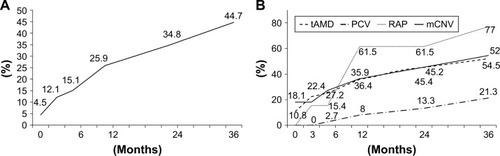
Table 2 Results of univariate analysis between the ORT(+) and ORT(−) groups