Figures & data
Table 1 Characteristics of eyes with RPD and those of the control
Figure 1 Subfoveal choroidal areas of EDI-OCT image in eyes with RPD and control eyes.
Notes: The choroidal, luminal, and stromal areas in eyes with RPD were significantly smaller than those of the control eyes (*P=0.001, unpaired t-test).
Abbreviations: EDI-OCT, enhanced depth imaging optical coherence tomography; RPD, reticular pseudodrusen.
Abbreviations: EDI-OCT, enhanced depth imaging optical coherence tomography; RPD, reticular pseudodrusen.
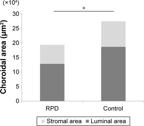
Table 2 Results of choroidal binarization to quantify the total, luminal, and stromal choroidal areas in patients with RPD and in control subjects
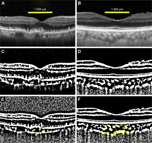
Figure 2 EDI-OCT images of an eye with RPD and a control eye.
Notes: The EDI-OCT image of the eye with RPD (A) was converted to a binary image (C) by using the ImageJ software. An EDI-OCT image of the control eye (B) was converted to a binary image (D). The choroidal area measured was 1,500 µm wide with the margins 750 µm nasal and 750 µm temporal to the fovea. Vertically, the area extended from the retinal pigment epithelium to the chorioscleral border. The yellow area is the measured area of the choroid (E and F).
Abbreviations: EDI-OCT, enhanced depth imaging optical coherence tomography; RPD, reticular pseudodrusen.
Abbreviations: EDI-OCT, enhanced depth imaging optical coherence tomography; RPD, reticular pseudodrusen.