Figures & data
Figure 1 RT-PCR products of LTA mRNA detected by 3% agarose gel electrophoresis stained by ethidium bromide.
Notes: mRNA expression of LTA was detected in the two peripheral blood samples from healthy subjects (lanes 1 and 2). Interestingly, RT-PCR analysis revealed different alternative LTA transcripts (A, B and C). Automatic sequencing revealed that transcript A retained introns 2 and 3 of LTA, transcript B retained intron 2, while transcript C lacked exon 3 of LTA. However, all of these three transcripts had altered reading frames and included premature stop codons, leading to truncated LTA isoforms that may have no function. GAPDH expression was used as internal control. Lane M was ΦX174 DNA/HaeIII molecular weight marker (Promega). Lane (−) was a control reaction lacking reverse transcriptase.
Abbreviations: RT-PCR, reverse transcription-polymerase chain reaction; LTA, lymphotoxin alpha; mRNA, messenger RNA.
Abbreviations: RT-PCR, reverse transcription-polymerase chain reaction; LTA, lymphotoxin alpha; mRNA, messenger RNA.
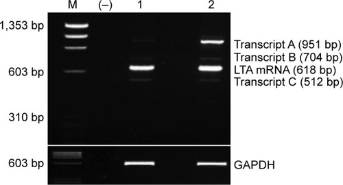
Figure 2 Expression analysis of T and C alleles of LTA rs2229094 polymorphism.
Notes: COS1 cells were transfected with plasmids encoding LTA-C allele (pCEFL-Flag-LTA-C) and LTA-T allele (pCEFL-Flag-LTA-T). (A) mRNA expression of both LTA alleles was assessed by RT-PCR at 7, 24 and 48 hours after transfection. GAPDH expression was used as internal control. Lane M was ΦX174 DNA/HaeIII molecular weight marker (Promega). Lane (−) was a control reaction lacking reverse transcriptase. (B) Protein expression of both LTA alleles was evaluated by Western blot at 48 hours after transfection using anti-Flag antibody. Lane 1 was LTA-C allele, lane 2 was LTA-T allele and lane (−) was a negative control (not transfected COS1 cells). β-Actin expression was used as internal control. No differences in RNA or protein expression levels were observed between the LTA-C and T alleles.
Abbreviations: LTA, lymphotoxin alpha; RT-PCR, reverse transcription-polymerase chain reaction.
Abbreviations: LTA, lymphotoxin alpha; RT-PCR, reverse transcription-polymerase chain reaction.
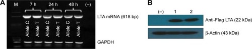
Figure 3 Subcellular localization of T and C alleles of LTA rs2229094 polymorphism.
Notes: COS1 cells were transfected with plasmids encoding LTA-C allele (pCEFL-Flag-LTAC) and LTA-T allele (pCEFL-Flag-LTAT). Forty-eight hours after transfection, confocal immunofluorescence assay was performed using anti-Flag antibody (green) and cell nuclei were stained with DAPI (blue). Both LTA-C allele (A) and T allele (B) were mainly located in the cytoplasm, without finding any differences in their cellular localization. (C) Not transfected COS1 cells.
Abbreviations: LTA, lymphotoxin alpha; DAPI, 4′,6-diamidino-2-phenylindole.
Abbreviations: LTA, lymphotoxin alpha; DAPI, 4′,6-diamidino-2-phenylindole.
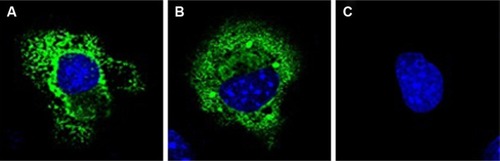
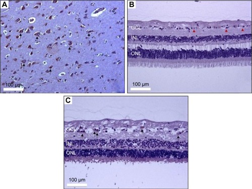
Figure 4 Immunohistochemical assay of LTA in human retina.
Notes: Immunohistochemical images of (A) adult human hippocampus anti-LTA staining, showing intense positivity (+++) within the cytoplasm of hippocampal neurons (black arrows), 20×. (B) Healthy human retina anti-LTA staining (control). Negative LTA cytoplasmic positivity (−) is observed in ganglion cells (arrowheads), 20×. (C) Anti-LTA staining (case) in human retina with chronic retinal detachment showing moderate cytoplasmic positivity (++) in ganglion cells (black arrows), 20×.
Abbreviations: LTA, lymphotoxin alpha; GCL, ganglion cell layer; INL, inner nuclear layer; ONL, outer nuclear layer.
Abbreviations: LTA, lymphotoxin alpha; GCL, ganglion cell layer; INL, inner nuclear layer; ONL, outer nuclear layer.