Figures & data
Figure 1 External view (top) and the schematic diagram of the cross-sectional view of the contact lens (bottom).
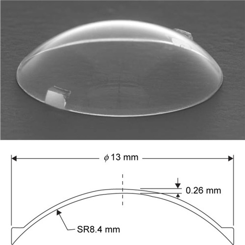
Figure 2 Fundus view of the contact lens with the noncontact wide-angle viewing system in a case of rhegmatogenous retinal detachment.
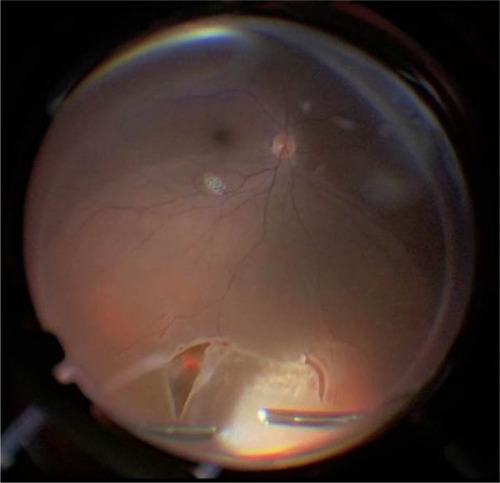
Figure 3 Fundus view of the wide-angle viewing system with a 128-diopter objective lens alone (top), and use of the contact lens with the wide-angle viewing system (bottom) for removal of proliferative membrane in a case of proliferative vitreoretinopathy (left) and for performing posterior vitreous separation in a case of vitreomacular traction (right).
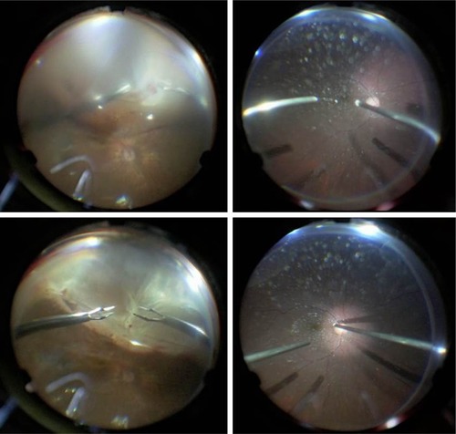
Figure 4 Fundus view through the contact lens with a 128-diopter objective lens (left) and with a 60-diopter objective lens (right) in a case of proliferative diabetic retinopathy.
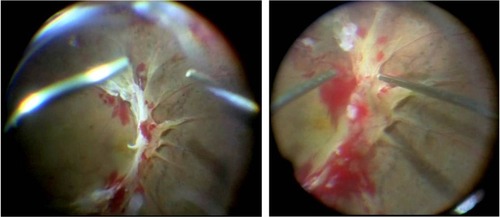
Figure 5 Application of the contact lens on the cornea in a case of diabetic retinopathy with corneal erosion and epithelial edema (top). Fundus view of the wide-angle viewing system alone (bottom left) and during use of the contact lens with the wide-angle viewing system (bottom right).
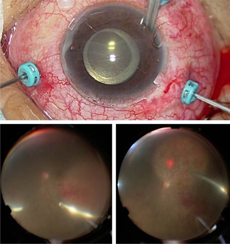
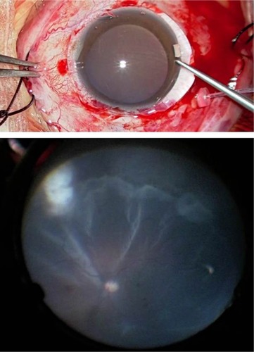
Figure 6 Application of the contact lens in a scleral buckling case (top). Fundus view through the contact lens with the wide-angle viewing system (bottom).