Figures & data
Figure 1 A) Affected eye at presentation with mild corneal edema and fine keratic precipitates. B) Scleritis with scleral thinning and subtenon’s triamcinolone.
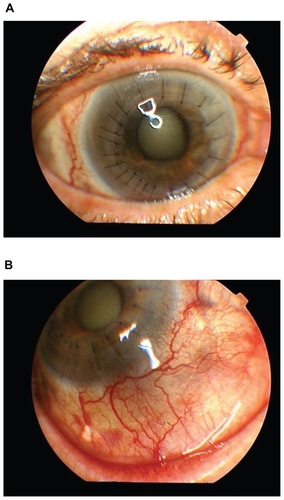
Figure 2 Confocal microscopy demonstrating cystic structures in the corneal stroma consistent with Acanthamoeba.
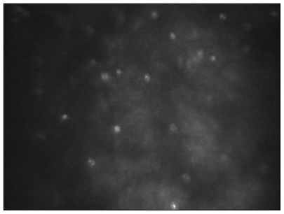
Figure 3 Stromal acanthamoebal cyst. PAS X 400.
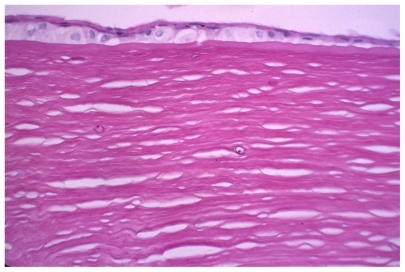
Figure 4 Loculated mass in superior anterior chamber with sclertitis.
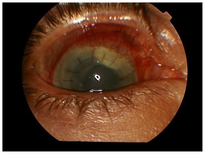
Figure 5 Confocal microscopy.
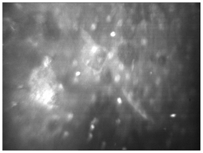
Figure 6 Acanthamoeba cysts within corneal stroma.
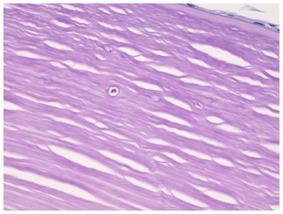
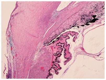
Figure 7 Inflammation in sclera, ciliary body, and choroid.