
Figures & data
Figure 1 High-definition optical coherence tomography at center of cornea.
Note: Top caliper measures flap thickness at 132 μm and bottom caliper measures residual stromal bed thickness at 346 μm.
Abbreviations: T, temporal; N, nasal.
Abbreviations: T, temporal; N, nasal.
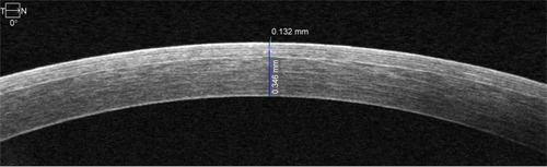
Figure 2 High-definition optical coherence tomography measurements.
Notes: Example of measurements across the cornea. Five measurements are spaced approximately 1.5 mm apart from each another, with the center measurement at the center of the cornea. Top calipers measure flap thickness. Bottom calipers measure residual stromal bed thickness.
Abbreviations: T, temporal; N, nasal.
Abbreviations: T, temporal; N, nasal.
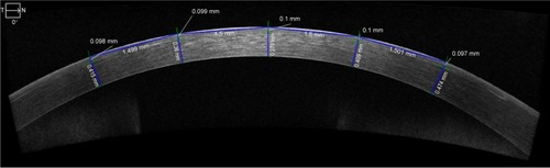
Table 1 Timing of OCT measurements
Table 2 Flap- and RSB-thickness comparison
Table 3 Summary of overall clinical outcome of LASIK patients