Figures & data
Figure 1 Photographs of a 81-year-old woman with retinitis pigmentosa. A) Fundus photographs show bilateral retinal arteriolar narrowing and intraretinal pigment deposition (bone spicule pigmentation) in both eyes, left: left eye, right: right eye. B) A fundus photograph of the right eye using a slit-lamp examination shows a detached retina in the nasal periphery. C) Fluorescein angiography images of the right eye show a hyperfluorescence due to atrophy of retinal pigment epithelium and hypofluorescence in the detached area.
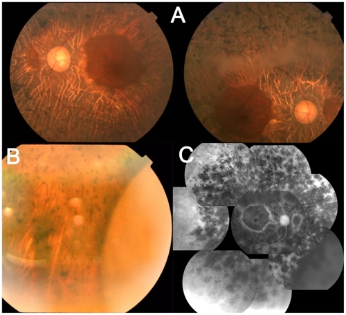
Figure 2 Visual fields of left eye (left) and right eye (right) of a patient with retinitis pigmentosa using a Goldmann perimeter were markedly constricted to the central 10° diameter.
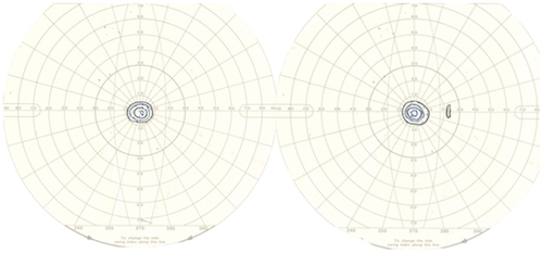
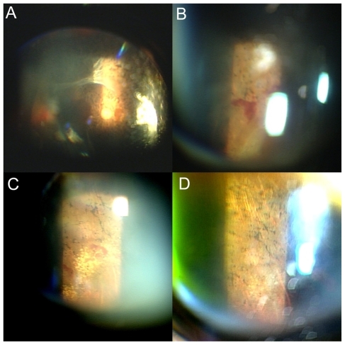
Figure 3 The right eye of the same patient. A) and B) A fundus photograph using a slit-lamp examination of 5 months later shows that the posterior vitreous detachment further progressed beyond the arcade area A), accompanying retinal hemorrhage around the detached retina B). C) A fundus photograph using a slit-lamp examination of 8 months later shows that the retina is reattached, and hard exudates are observed in the nasal periphery. D) A fundus photograph using a slit-lamp examination of 12 months later shows that those hard exudates are resolved.