
Figures & data
Table 1 Modification of Donnenfeld nomogram
Figure 1 Basic procedure: place incisions, (A) align suction ring, (B) set parameters, (C and D) confirm 3D depth with optical coherence tomography.
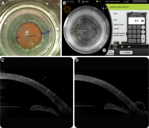
Table 2 Preoperative Kcyl, postoperative Kcyl, and postoperative RRA (in diopters)
Figure 2 Percentage of eyes with ≤0.5 diopters of keratometric cylinder preoperative and postoperative in eyes with against-the-rule, oblique, and with-the-rule astigmatism, and all astigmatic eyes combined.
Abbreviations: ATR, against-the-rule; OBL, oblique; WTR, with-the-rule.
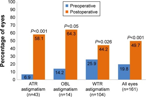
Figure 3 Correction index vector plots, preoperative Kcyl and postoperative Kcyl.
Abbreviations: Kcyl, keratometric corneal cylinder; WTR, with-the-rule; ATR, against-the-rule; OBL, oblique.
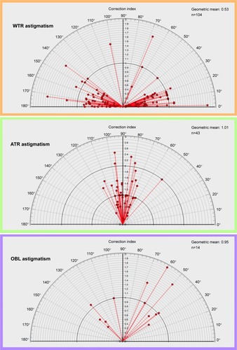
Table 3 Distribution of overcorrected eyes after femtosecond-assisted arcuate incisions