
Figures & data
Table 1 Primary antibodies and incubation protocols
Figure 1 Whole tissue section immunohistochemistry.
Notes: (A) Neuron-specific enolase. In this case, there was widespread and intense staining for neuron-specific enolase. 10×. (B) Synaptophysin. This RB sample also had widespread and intense staining for synaptophysin. 20×. (C) Synaptophysin distribution in Flexner–Wintersteiner rosettes. This RB had greater staining intensity for synaptophysin located in the apical cytoplasm of Flexner–Wintersteiner rosettes. 40×. (D) pRb. This was diffusely present in the nuclei of RB cells. 40×. (E) RB negative for pRb. The tumor cells in this RB did not express pRb; however, the vascular endothelial cells, which served as an internal immunohistochemical control, did express it. 40×.
Abbreviations: pRb, RB-associated protein; RB, retinoblastoma.
Abbreviations: pRb, RB-associated protein; RB, retinoblastoma.
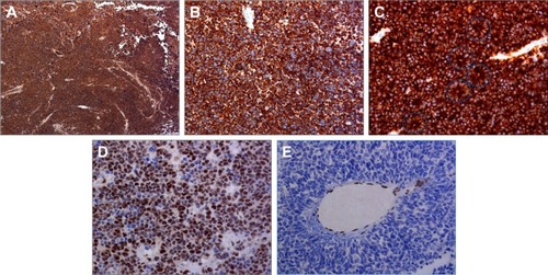
Table 2 Immunohistochemistry: whole tissue sections
Table 3 Immunohistochemistry: whole tissue sections
Table 4 Immunohistochemistry: neuron-specific enolase: whole tissue sections and tissue microarray
Figure 2 Tissue microarray immunohistochemistry.
Notes: (A) Neuron-specific enolase. This case shows widespread and intense staining for neuron-specific enolase. 5×. (B) Synaptophysin. All tumor cells present in the core were intensely positive for synaptophysin. 5×. (C) pRb. All of the nuclei were positive for pRb. 40×.
Abbreviation: pRb, retinoblastoma-associated protein.
Abbreviation: pRb, retinoblastoma-associated protein.
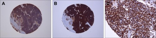
Table 5 Immunohistochemistry: synaptophysin: whole tissue sections and tissue microarray
Table 6 Immunohistochemistry: pRb: whole tissue sections and tissue microarray