
Figures & data
Figure 1 Myopia progression rate (D/month) in both groups.
Notes: This rate is significantly higher in the MH group compared with the ERM group (0.42±0.25 vs 0.29±0.19, P=0.035). *Unpaired t-test.
Abbreviations: MH, macular hole; ERM, epiretinal membrane.
Abbreviations: MH, macular hole; ERM, epiretinal membrane.
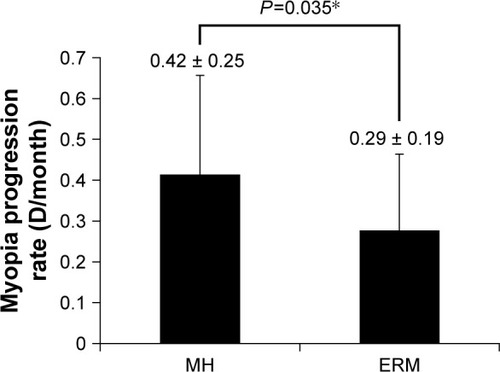
Table 1 Preoperative, intraoperative, and postoperative factors in both groups
Table 2 Receiving cataract surgery after vitrectomy in both groups and tamponade material in MH group
Figure 2 Correlation between myopia progression rate and four factors (surgical time, gender, TA use, and BBG use) in the MH group.
Notes: Correlation with surgical time is plotted in (A). Comparison to gender rate is shown in (B) (male; 0.14±0.12 vs female; 0.46±0.25, P<0.001). Comparison of TA use and BBG use is shown in (C) and (D), respectively. *Unpaired t-test, ***Pearson’s correlation coefficient, #impossible to compare.
Abbreviations: TA, triamcinolone acetonide; BBG, brilliant blue G; MH, macular hole.
Abbreviations: TA, triamcinolone acetonide; BBG, brilliant blue G; MH, macular hole.
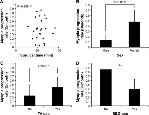
Table 3 Correlation coefficients of factors related to myopia progression rate in the MH group
Table 4 The mean age of patients in the MH group