Figures & data
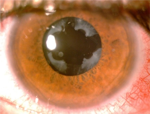
Figure 1 Slit lamp photograph showing signs of cataract and pseudoexfoliation.
Notes: The lens surface shows sign of exfoliating fibrillar material with accumulation of whitish flakes at the pupillary margin. The pupil is only moderately dilated after pharmacological mydriasis.
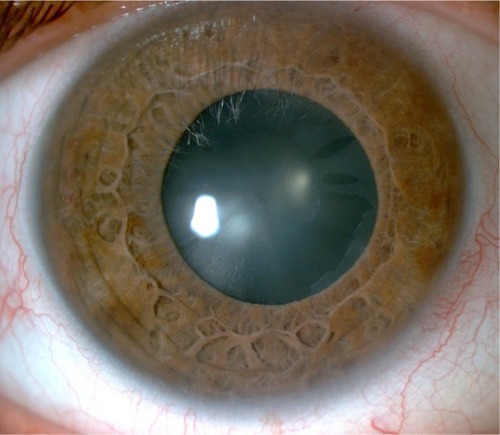
Table 1 Preoperative risk factors and intraoperative and postoperative complications in PXF patients with cataract surgery
Figure 2 Intraoperative photograph showing capsule retractors stenting the capsular bag during phacoemulsification.
Note: Following cortical lens aspiration, a capsular tension ring is implanted prior to intraocular lens implantation.
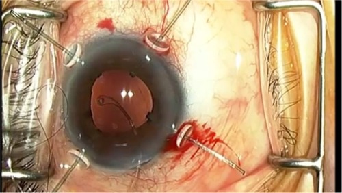
Figure 3 Slit lamp photograph showing anterior capsule opacification and phimosis after cataract surgery with in-the-bag IOL implantation.
Note: Note the reduction in size of the anterior capsulotomy with radial lines of capsular shrinkage, indicating traction on the peripheral lens zonules.
Abbreviation: IOL, intraocular lens.
Abbreviation: IOL, intraocular lens.
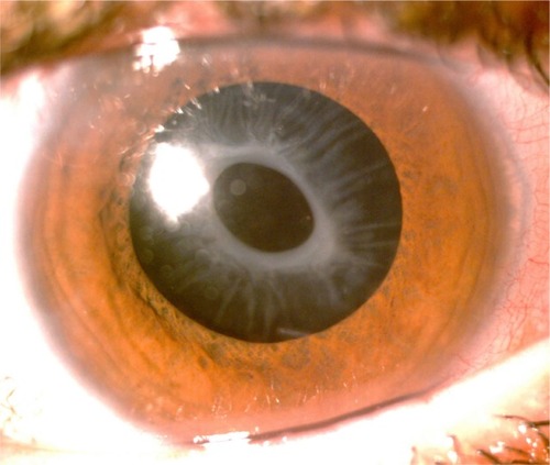
Figure 4 Slit lamp photograph after Nd:YAG laser anterior capsulotomy.
Note: Lens capsule contraction was released with enlargement of the anterior capsulotomy.