Figures & data
Table 1 Baseline patient characteristics
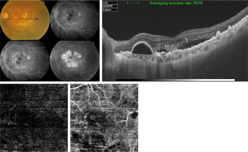
Figure 1 Upper left, color photo and FFA of the right eye of a 55-year-old male with exudative AMD and classic CNV. Upper right, corresponding SS-OCT image in radial scan mode shows hyper-reflective nodular lesion located entirely above the RPE. The lesion resulted in disruption of the retinal layers and is associated with gross intra-retinal edema which indicates type II active CNV. Lower left, “en face” SS-OCTA image of the same eye taken at the level of the outer retina in a 3×3 mm field. The neovascular complex is displayed as a hyperintense signal caused by increased blood flow within the lesion. The remaining avascular outer retina generates a hypointense signal due to absent blood flow and is displayed as a dark-gray background. The lesion demonstrates homogenous network of tiny interlacing capillaries with occasional larger vessels displayed as jet-white streaks (closed arrow heads). The entire complex is surrounded by a dark lucid interval intervening between the lesion and the surrounding normal outer retina (open arrow heads).
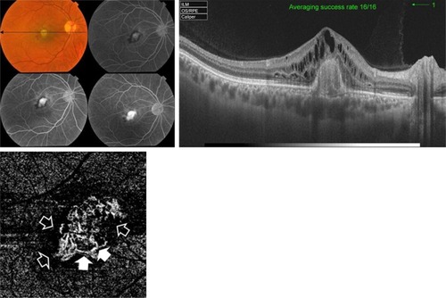
Figure 2 Upper left, color photo, red-free photo, and FFA of the right eye of a 51-year-old female with classic CNV on top of MFC. Upper right, corresponding SS-OCT image in line scan mode shows three hyper-reflective nodular lesions located entirely above the RPE. The sub-foveal lesion resulted in disruption of the outer retinal layers and is associated with overlying intra-retinal edema which indicates type II active CNV. Lower left, “en face” SS-OCTA image of the same eye taken at the level of the outer retina in a 3×3 mm field. The central portion of the lesion demonstrates dense arborization. More peripherally, several vascular anastomosis (closed arrow heads) and looping (open arrow head) are shown.
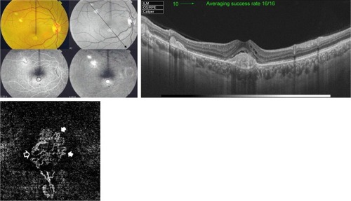
Figure 3 Upper left, color photo and FFA of the left eye of a 75-year-old female with scarred CNV secondary to AMD. Upper right, corresponding SS-OCT image in radial scan mode shows marked disorganization of the outer retina with marked thinning of the RPE and choriocapillaris. No intra-retinal fluid is discernible. Lower, “en face” SS-OCTA image of the same eye taken at the level of the outer retina (left) and the choriocapillaris (right) in a 6×6 mm field. The entire lesion complex is composed mostly of large mature vessels that yield hyperintense signal with intervening hollowness due to absent capillaries. A single large feeder vessel is shown supplying the main lesion (closed arrow heads). The shadow of the large choroidal vessel (open arrow head) is shown.
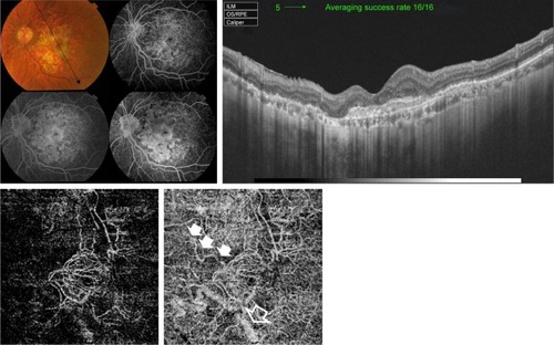
Figure 4 Upper, SS-OCT of the left eye of an 80-year-old male with AMD. Marked disorganization of the outer retina with marked thinning of the RPE and choriocapillaris and minimal intra-retinal fluid accumulation are shown. Lower, corresponding “en face” SS-OCTA image taken at the level of the outer retina (left) and the choriocapillaris (right) in a 6×6 mm field. The medusa head configuration composed of at least five large mature vessels (arrows) emanating from single main trunk (arrow head) is shown.
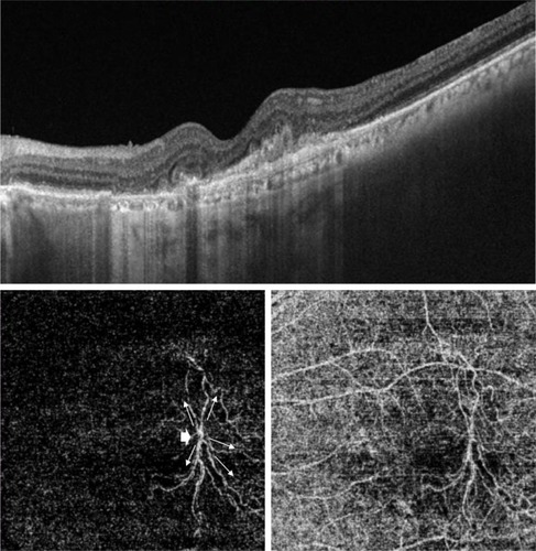
Figure 5 Upper, SS-OCT image of the left eye of a 66-year-old female with neovascular AMD shows multiple RPE detachments. The RPE in the sub-foveal area is irregular, thickened, and raised-up by a moderately hyper-reflective lesion associated with disorganization of the outer retina and accumulation of sub- and intra-retinal fluid, which indicates type I CNV. Lower, “en face” SS-OCTA image captured at the level of the outer retina (left) and choriocapillaris (right) in a 3×3 mm field. The extent of arborization of the neovascular complex is most revealed at the choriocapillaris projection (closed arrow head) compared to the outer retina projection (open arrow head).
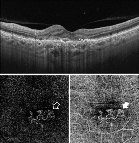
Figure 6 Upper, SS-OCT of the left eye of a 30-year-old female with MFC shows hyper-reflective double hump lesion causing disorganization of the outer retina and is associated with intra-retinal fluid. The lesion is located entirely above the RPE, which indicates type II CNV. Lower, “en face” SS-OCTA image captured at the level of the outer retina (left) and choriocapillaris (right) in a 3×3 mm field. The outer boundaries of the lesion and the arborizing pattern of the neovascular network are best delineated in the outer retina projection, whereas in the choriocapillaris projection the boundaries of the lesion are indistinguishable from the surrounding background in several locations.
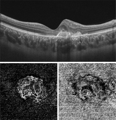
Figure 7 Upper left, color photo and FFA of the left eye of a 32-year-old female with high myopia and sub-foveal hemorrhage. The blocked fluorescence on FFA in the foveal region is shown. Upper right, corresponding SS-OCT image in a radial scan mode shows hyper-reflective sub-foveal lesion corresponding to the hemorrhage seen in color photo and FFA. Both, FFA and SS-OCT are inconclusive for CNV development. Lower, “en face” SS-OCTA image captured at the level of the outer retina in a 6×6 mm field (left) and co-registered OCT segmentation slabs (right). There is no identifiable decorrelation signal characteristic of abnormal vascular network formation in the area of suspicious CNV formation.
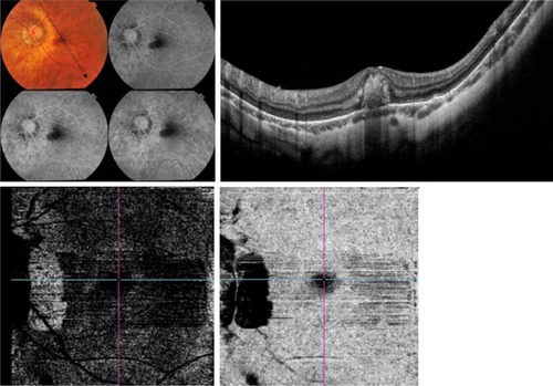
Figure 8 Upper left, color photo and FFA of the left eye of a 66-year-old female with intermediate AMD and extensive drusen. The late frames of FFA show pooling in multiple PEDs and LLIO characteristic of occult CNV. Upper right, corresponding SS-OCT image in radial scan mode shows RPE detachment. The RPE is irregular, thickened, and raised-up by a moderately hyper-reflective lesion with accumulation of sub-retinal fluid, suggestive of type I CNV formation. Lower, “en face” SS-OCTA image of the same eye taken at the level of the outer retina in a 3×3 mm field (left) and co-registered OCT segmentation slabs (right). The absence of identifiable decorrelation signal characteristic of abnormal vascular network formation in the area of suspicious CNV formation is shown.