Figures & data
Figure 1 (A) Specular microscopy from a normal cornea. (B) Specular microscopy from a patient with iridocorneal endothelial syndrome.
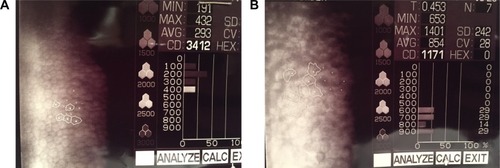
Figure 2 Xen45 stent (AqueSys Inc., Aliso Viejo, CA, USA) visible in the conjunctival space in a patient with iridocorneal endothelial syndrome.
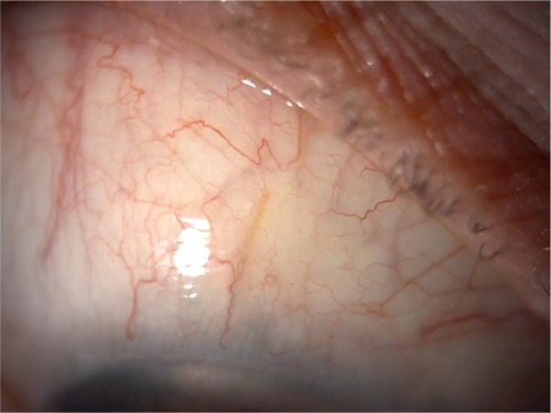
Figure 3 Broad peripheral anterial synechiae seen in iridocorneal endothelial syndrome.
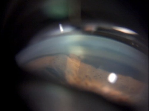
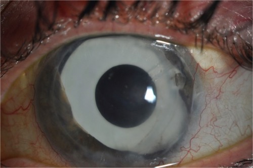
Figure 4 A patient who has undergone multiple procedures for iridocorneal endothelial syndrome. A clear Descemet stripping automated endothelial keratoplasty graft is visible, with a supertemporal Baerveldt tube in situ. A prosthetic iris implant can also be seen.