Figures & data
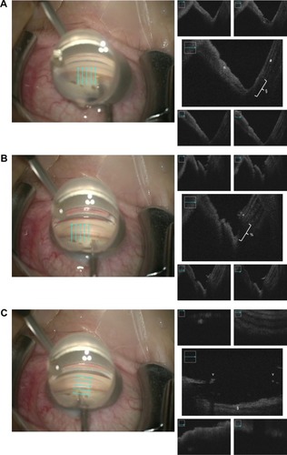
Figure 1 Color images: view from above through the OPMI LUMERA 700 surgical microscope (Carl Zeiss Meditec AG, Jena, Germany). Cyan arrows indicate the scan locations of the iOCT. Black and white images: corresponding iOCT scans of the ACA. (A) At the beginning of the surgery: *iris and §trabecular meshwork. (B) Postprocedure: >anterior part of the trabecular meshwork opening and %area of Schlemm’s canal. (C) Postprocedure: &blood blocks the signal in the area of the trabecular meshwork opening.
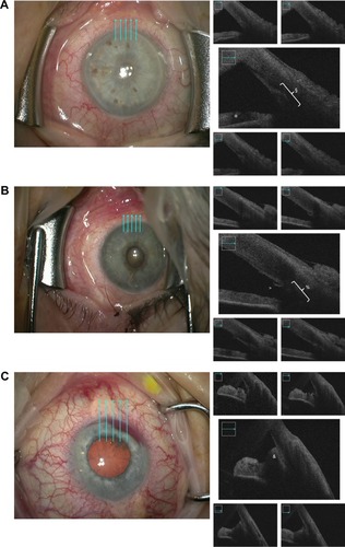
Figure 2 Color images: OPMI LUMERA 700 surgical microscope (Carl Zeiss Meditec AG, Jena, Germany) view through modified Swan-Jacob goniolens (Ocular Instruments, Bellevue, WA, USA) into the ACA. Cyan arrows indicate scan locations of the iOCT. Black and white images: corresponding iOCT scans of the ACA. (A) At the beginning of the surgery: *iris, §trabecular meshwork, and #cornea. (B) Postprocedure, vertical scans: <anterior part of the trabecular meshwork opening and %area of Schlemm’s canal. (C) Postprocedure, horizontal scans: vborder of the trabecular meshwork opening and $scleral backwall of Schlemm’s canal.