Figures & data
Figure 1 Auto-peeling of idiopathic ERM can be seen in these fundus photos and macular OCT from a 33-year-old female with idiopathic ERM. These photos and OCT were taken in (A) July 2010 and (B) October 2012. Visual acuity was improved from 10/20 to 14/20.
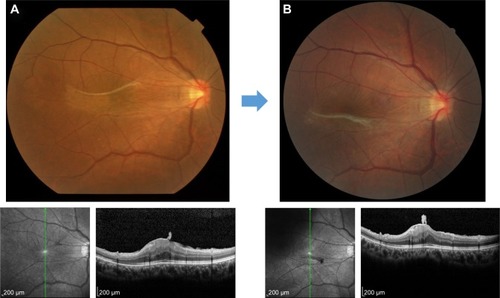
Figure 2 Auto-peeling of idiopathic ERM can be seen in these fundus photos from a 76-year-old female with idiopathic ERM. These photos were taken in (A) August 2008, (B) November 2009, and (C) February 2012.
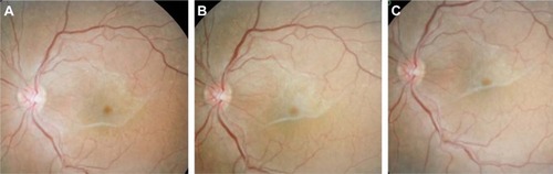
Figure 3 Recurrence of ERM in the macula after successful removal of idiopathic ERM by vitrectomy. Vitrectomy for idiopathic ERM was performed and visual acuity improved from 20/30 (A) to 20/20 (B). However, it reoccurred 1 year after the vitrectomy and the visual acuity decreased to 20/30 (C).
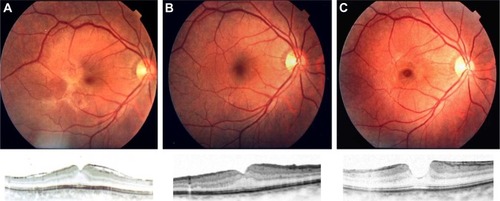
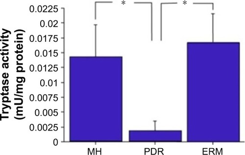
Figure 4 Tryptase activity in the vitreous. Tryptase activity in the vitreous was significantly higher in patients with ERM (n=14) and MH (n=14) compared to PDR (n=13); *P<0.05.