
Figures & data
Figure 1 Procedure of ophthalmic endoscope-guided subretinal fluid drainage in pars plana vitrectomy for RRD.
Notes: (A) First, the patient’s head is moved toward the position where the primary retinal break is located at the lowest level. In this position, subretinal fluid can easily come out from the retinal break into the vitreous space. (B) We mainly inserted the buckflash needle with a silicone tip (a) from the higher positioned port and the ophthalmic endoscopic probe (b) from the lower positioned port. But we used these instruments oppositely depending on the case or situation.
Abbreviation: RRD, rhegmatogenous retinal detachment.
Abbreviation: RRD, rhegmatogenous retinal detachment.
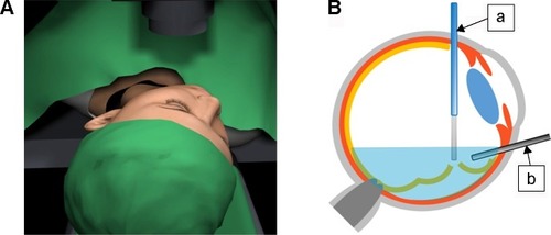
Figure 2 Representative images under the 25-gauge ophthalmic endoscope.
Notes: (A) Subretinal fluid drainage was performed through a primary retinal break during fluid–air exchange. (B) Endolaser photocoagulation was performed around the primary retinal break. (C) Small retinal tear was found at peripheral retina (arrow) during fundus inspection under the air condition. (D) Performing endolaser photocoagulation around the small retinal tear.
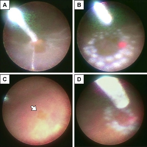
Table 1 Characteristics of 127 eyes of 127 patients who underwent endoscope-assisted vitrectomy
Table 2 Overall and comparison of postoperative success rates between 23- and 25-gauge group
Table 3 Visual acuity outcomes
Table 4 Comparison of surgical time between 23- and 25-gauge group in different surgical procedures
Table 5 Overall intraoperative and postoperative complication rates and comparison between 23- and 25-gauge group